


Chiba Medical J. 93E:39~43,2017
doi:10.20776/S03035476-93E-4-P39
[ Case Report ]
Tomotaka Umimura1), Kazuki Fujimoto1), Sumihisa Orita1),
Hiroto Kamoda2), Kazuyo Yamauchi1), Miyako Suzuki1),
Kazuhide Inage1),
Jun Sato3), Yasuhiro Shiga1),Koki Abe1),
Hirohito Kanamoto1), Masahiro Inoue1), Hideyuki Kinoshita1),
Masao Koda1), Takeo Furuya1), Kazuhisa Takahashi1),
and Seiji Ohtori1)
1) Department of Orthopaedic Surgery, Graduate School of Medicine, Chiba University, Chiba 260-8670.
2) Department of Orthopaedic Surgery, Chiba Cancer Center, Chiba 260-8717.
3) Department of Orthopaedic Surgery, Chiba Aoba Municipal Hospital, Chiba 260-0852.
(Received January 26, 2017, Accepted March 13, 2017)
Introduction: Total Spondylectomy(TS) of L5 remains challenging given its anatomical position.
The reconstruction materials have not been designed for L5 reconstruction and shaping them to fit the lumbar lordosis is difficult. An expandable cage is the most reliable implant for reconstruction of the anterior column.
Case presentation: A 68-year-old man presented with a high grade sarcoma of an L5 lumbar vertebra, which we treated surgically using a two-stage TS. At the time of admission, the patient complained of lower extremity pain due to spinal nerve root compression and he had difficulty walking. We performed TS, and acquired three-column stability using a wedge resection of the S1 endplate, an expandable cage, and fixation using pedicle screws and rods. His lower extremity pain improved and he could walk unassisted 6 months after surgery.
Conclusion: When TS of L5 is required, an expandable cage may be useful for reconstructing the anterior column. We further propose that TS of L5, should be considered a two-stage surgery involving a combination of posterior and anterior approaches, performed on separate days. Such an approach is recommended because of the complexity of the anatomical position, the biomechanical reconstruction difficulty, and the invasiveness of the surgery.
total spondylectomy, spinal tumor, surgery, anterior reconstruction,
expandable cage
The goals of surgical treatment of lumbosacral malignancy are to relieve dura or nerve compression, remove malignant tissue, and reconstruct a stable spine. In recent years, TS surgery has become more prevalent as a curative treatment approach for metastatic spinal tumors and for primary spinal malignancies. However, TS of the lower lumbar spine, particularly at L5, remains challenging given its anatomical position(major vessels bifurcate in front of the vertebral body and nerve roots are located behind the vertebral body) and biomechanical properties[1]. We report a patient who underwent TS for a primary, malignant, L5 spinal tumor.
A 68-year-old man reported to his local hospital with a complaint of lower back and leg pain. X-ray and computed tomography(CT) images revealed a lumbar spinal tumor at L5, and he was referred to our hospital. An excisional biopsy of the tumor revealed a low-grade sarcoma. At the time, he had difficulty walking because of the severe bilateral leg pain. A physical examination demonstrated motor weakness of the extensor hallucis longus(Manual Muscle Test, 3/5). He had no bladder or rectal disturbances. Sensory examination confirmed hypoalgesia on both sides of the L5 distribution area (Figure 1). Laboratory investigation showed that C-reactive protein(CRP) levels were slightly elevated (3.5), and enzyme assays indicated that his liver and renal functions were within normal ranges.
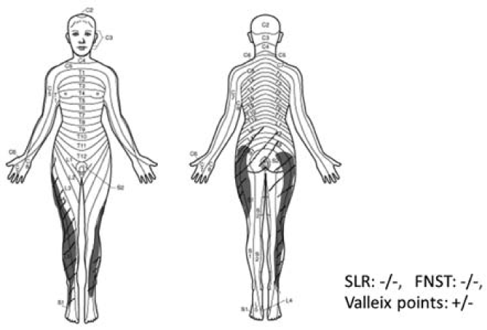
Fig. 1 Neurologic findings at the time of admission
Sensory examination confirmed bilateral hypoalgesia of the L5 distribution area and motor weakness of the extensor hallucis longus(Manual Muscle Test, 3/5). The patient had no bladder or rectal disturbances. Straight leg raise(SLR) test and femoral nerve stretch test(FNST) were both negative.
X-ray and CT imaging showed that most of the L5 vertebral body had been destroyed by the tumor. Magnetic resonance imaging further revealed that the tumor had invaded into most of the L5 vertebral body. The tumor had also invaded the intervertebral foramen, severely compressing the L5 nerve roots, bilaterally (Figure 2). In CT angiography, the bifurcation of the major vessels was located in front of the L5 vertebral body(Figure 3).
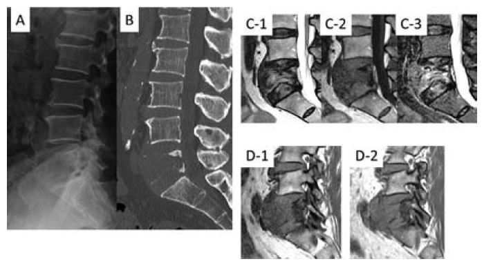
Fig. 2 Imaging findings at the time of admission
X-ray(A) and sagittal computed tomography(B) images demonstrate that most of the L5 vertebral body was destroyed. Sagittal T1-weighted(C-1), T2-weighted(C-2), short tau inversion recovery sequence(C-3) magnetic resonance images demonstrate that the tumor had invaded most of the L5 vertebral body. Sagittal T1-weighted images of the bilateral intervertebral foramen(D-1; Right side,D-2; Left side) demonstrate that the tumor had severely compressed the L5 nerve roots, bilaterally.
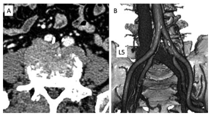
Fig. 3 Computed tomography angiography imaging studies
Axial(A) and 3-dimensional(B) images show the bifurcation of the descending aorta and inferior vena cava in front of the L5 vertebral body.
We decided to treat this tumor with TS as a curative surgery. We planned a TS with spinal instrumentation via a posterior-anterior approach.
Surgery was performed in two stages on the same day:(1) posterior TS and spinal reconstruction, and(2) anterior dissection around the vertebral body and the tumor, with anterior fusion.
Step 1 . We placed the patient in a prone position and made a posterior midline incision. We exposed the posterior elements from L3 to the sacrum; inserted screws into the bilateral pedicles of L3-S1(except for L5) and the bilateral ilium; and connected a rod to the bilateral screws, including the iliac screws. We removed the L5 vertebral arch, pedicles, and transverse process, and performed bilateral decompression of the L5 nerve roots. We stopped bleeding from the venous plexus in the spinal canal using a surgical cotton tamponade. We then washed the surgical site with saline, inserted a drain, and closed the wound.
Step 2 . For this stage, the patient was placed in a supine position and a transperitoneal approach was attempted via a medial abdominal incision. The descending aorta and inferior vena cava were dissected. This procedure was performed by vascular surgeons due to the technical difficulties caused by the bifurcation of both vessels in front of the tumor, and their apparent adherence to the tumor. Next, we performed dissection around the vertebral body. After detaching the L4/5 and L5/S discs, we resected most of the L5 vertebral body piece by piece, not en bloc, because of its fragility.
We had to make a big lordosis angle of about 40 degrees for reconstructing the anterior column, based on preoperative measurement. Instead, we performed 20-degrees partial wedge resection of the S1 head side endplate for inserting a 16-degree lordotic expandable cage for stability. Finally, we fixed it in place. The total surgical time, for both stages, was 11 hours and 23 minutes, with a total blood loss of 1750 mL(Figure 4).
The pathological diagnosis was a high-grade malignant sarcoma, in contrast to the preoperative diagnosis.
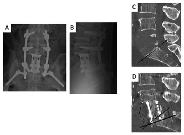
Fig. 4 Postoperative imaging studies
Anteroposterior(A) and lateral(B) x-ray images show the inserted expandable cage, a 16-degree lordotic cage, fixed in place. Preoperative(C) and postoperative(D) sagittal computed tomography images demonstrate the changes after 20-degrees partial wedge resection of the S1 head side endplate. The dotted line shows the angle of the S1 head side end plate before the operation. The black line indicates the angle of the endplate of the S1 head side after surgery.
Postoperatively, the patient’s condition was stable, without cage dislocation or alignment changes. Additionally, the patient’s lower extremity pain improved and he could engage in walker-assisted walking after 3 weeks, cane-assisted walking after 3 months, and unassisted walking after 6 months. However, he subsequently died of pulmonary metastasis after 7 months.
A 68-year-old man presented with a sarcoma of the L5 vertebral body, and underwent TS via combined posterior and anterior approaches. We could resect almost the entire tumor, and the patient’s symptoms improved markedly within 6 months after surgery.
Primary sarcomas of the spine are considered rare; they include osteosarcoma, chondrosarcoma, chordoma, and Ewing’s sarcoma. The average survival period has been reported as follows: chordoma, 96 months; Ewing’s sarcoma, 90 months; chondrosarcoma, 88 months; and osteosarcoma, 18 months[2]. Surgical resection plays an important part of curative treatment when these are single lesions, without metastasis, since they often respond poorly to either chemotherapy or radiation[3].
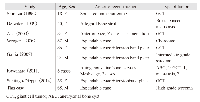
It was reported that TS can only be performed via a posterior approach for the upper lumbar spine. However, for the lower lumbar spine, a combined approach, such as a posterior-anterior or posterior-lateral approach, is recommended to avoid injury to the major vessels or to the nerve roots[4]. There have been numerous reports of TS at the L1-L4 levels, but few reports have described TS at L5. Only 12 cases of TS at L5 have been reported, to date; however, only two of these involved sarcomas(Table 1)[1,5-13].
Reported cases of total en bloc spondylectomy at L5
The reconstruction material used in the anterior column during TS surgery of the lumbar spine has been autologous iliac bone[14-16], autologous fibula[17], or a mesh cage[8]. However, these materials have not been designed for L5 reconstruction and shaping them to fit the lumbar lordosis is difficult. Furthermore, if the reconstruction does not fit well, it results in insufficient initial fixation.
On the other hand, an expandable cage is the most reliable implant for reconstruction of the anterior column as it can be inserted at a lordotic degree during attachment and it crimps easily[9]. However, the use of an expandable cage, alone, has been reported to give inadequate fixation force, based on cadaver studies [18]. There has been only one report of good results obtained using only an expandable cage[9]. Using an additional anterior lumbosacral plate seems to be necessary to obtain adequate stability, according to some reports [19,20]. Nevertheless, since an anterior lumbosacral plate cannot be used with the applicable health insurance in Japan, another method is needed to obtain the required stability. In our case, three-column stability was achieved using wedge resection of the S1 endplate, an expandable cage, and fixation using pedicle screws and rods.
We propose that TS of the lower lumbar spine, particularly at L5, should be considered a two-stage surgery, using a combination of posterior and anterior approaches. Further, we recommend performing the two stages on separate days because of the procedure’s technical difficulty. When using an expandable cage as the sole anterior reconstruction material, wedge resection of the lower vertebral endplate is important to prevent cage dislocation. This approach helps to reduce the invasiveness of the surgery.
We would like to thank the staff and management at the Department of Orthopedic Surgery, Graduate School of Medicine Chiba University. All of the authors, has no fund to declare in the present case.
Address correspondence to Dr. Kazuki Fujimoto.
Department of Orthopaedic Surgery, Graduate School of Medicine, Chiba University, 1-8-1, Inohana, Chuo-ku, Chiba 260-8670, Japan.
Phone: +81-43-226-2117. Fax: +81-43-226-2116.
E-mail:s9082@nms.ac.jp
Abbreviations: TS: total spondylectomy, CT: computed tomography, CRP: C-reactive protein
