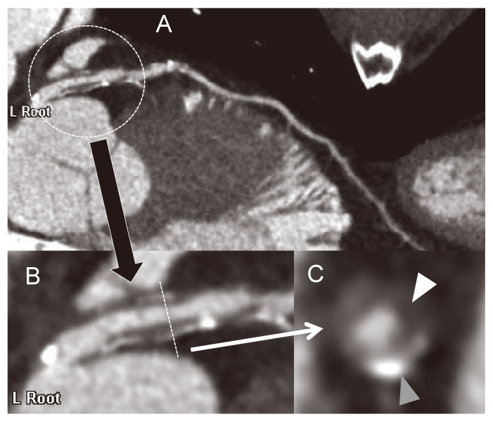
Fig. 1
Typical plaque image with positive remodeling, spotty calcification, and low attenuated lesion of a coronary artery in a curved planner reconstructed image on CT(A). The plaque image is magnified (B), and spotty calcification(gray arrow heads) and low attenuated plaque(white arrow heads) were obvious in a short axial image(C).