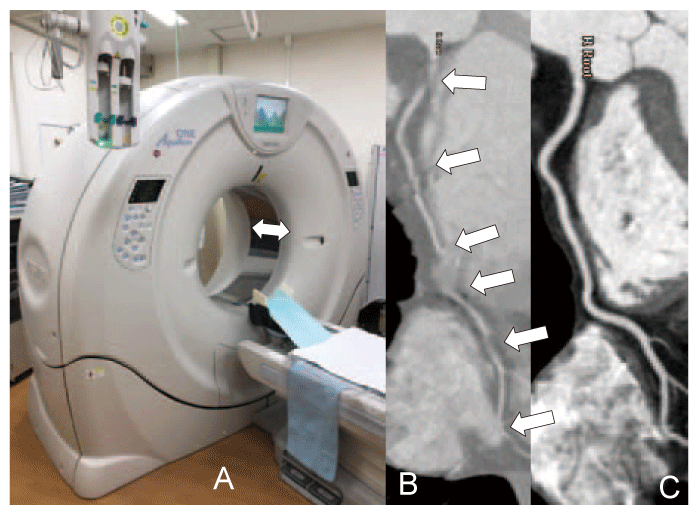
Fig. 2
320-slice Computed Tomography(CT)has a 16- cm craniocaudal coverage, which is indicated by a white arrow(A) , and it allows us to obtain clear images of coronary arteries without any stepping artifacts even in patients with frequent arrhythmia. Two typical different curved planner reconstructed images of the right coronary artery on CT in the same patient with atrial fibrillation on 16-slice CT (B)and 320-slice CT(C) . Stepping artifact was obvious on 16-slice CT(white arrows)because of its short craniocaudal coverage; however, the artifacts were diminished on 320-slice CT.