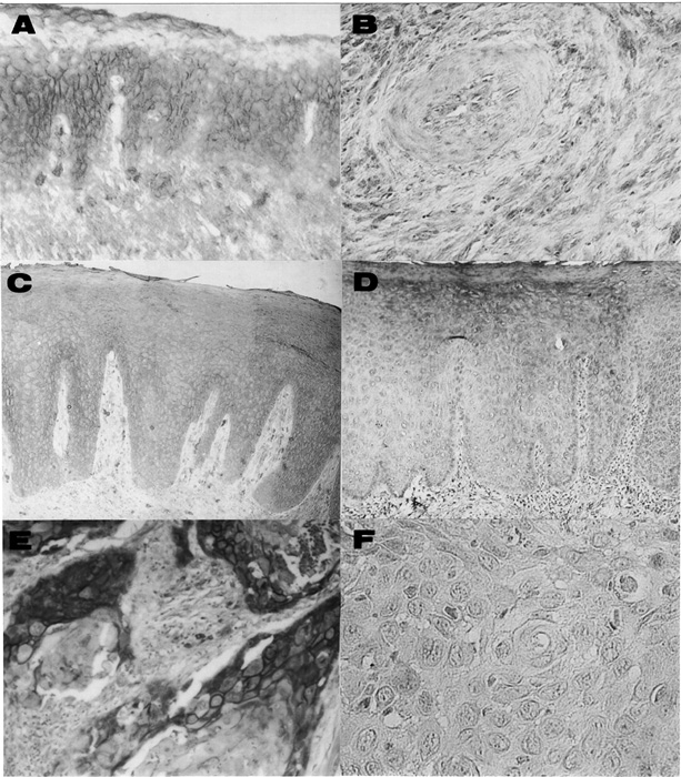
Fig. 1
Immunohistochemical staining of KAI1 in normal and cancerous tongue tissues.
- (A) Normal tongue tissue exhibited strong KAI1 protein expression that was limited to the cell membrane. Original magnification, X200.
- (B) KAI1-negative case of metastatic SCC. Original magnification, X400.
- (C) KAI1-positive case of leukoplakia. Note that strong positive immunoreaction for KAI1 was detected on the epithelial cell membrane. Original magnification, X200.
- (D) KAI1-negative case of leukoplakia. Original magnification, X200.
- (E) Positive staining of tumor cells of the primary tongue SCC. Original magnification, X400.
- (F) Negative staining of tumor cells of the primary tongue SCC. Original magnification, X400.