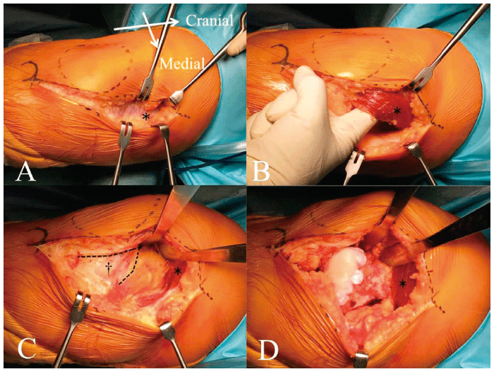
Fig. 1 Surgical exposure using the direct medial approach to the right knee
(A)A medial oblique incision of 14 cm is made from the medial aspect of the tibial tuberosity along the midpoint of the muscle belly of the vastus medialis obliquus muscle(*).(B)The muscle fibers are intentionally liftedup and lateralized within the sheath by the surgeon’s index finger.(C)The synovial layer of the suprapatellar pouch is visualized in a V-shaped flap(†).(D)The patella is lateralized with two narrow Homann retractors.
