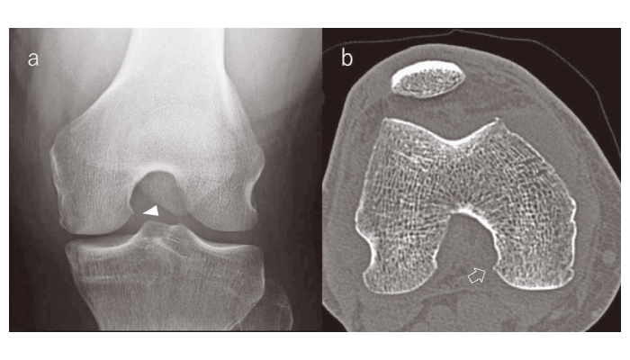
Fig.1 Representative CT and tunnel view to detect HOPOX
The right knee of a sixty-five year-old male was examined using x-rays (a) and CT (b) . The tunnel view revealed HOPOX formation (1a, arrowhead) on the medial side that was confirmed by CT (1b, open arrow)