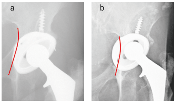
Fig.1 Case 1. Anteroposterior radiographs of the left hip at 5(a)and 20(b)years after the primary surgery. Polyethylene wear progressed and the cup gradually protruded into a defect in the medial wall of the pelvis(red line).
Fig.1 Case 1. Anteroposterior radiographs of the left hip at 5(a)and 20(b)years after the primary surgery. Polyethylene wear progressed and the cup gradually protruded into a defect in the medial wall of the pelvis(red line).