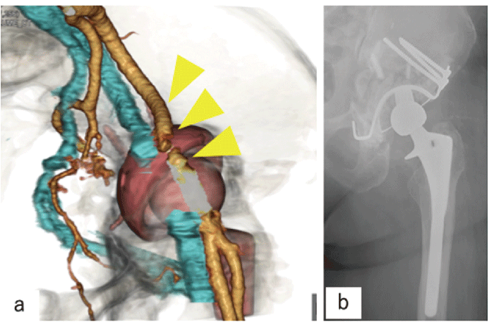
Fig.6 Case 3. Anteromedial view of the left pelvis on 3D CT angiography. The external iliac artery(beige line)and external iliac vein(blue line)were compressed and shifted superiorly by the edge of the migrated cup(yellow arrowhead). b. Postoperative anteroposterior left hip radiographs.