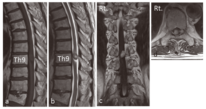
Fig. 2 Preoperative Radiological images. a, Preoperative T-1 weighted midsagittal magnetic resonance(MR)image. b, Preoperative T-2 weighted midsagittal MR image. c, Preoperative T-1 weighted after Gd-DTPA injection coronal MR image. d, Preoperative T-1 weighted after Gd-DTPA injection axial MR image showed a space-occupying lesion in the vertebral canal located on the middle to left side at the Th9 level.