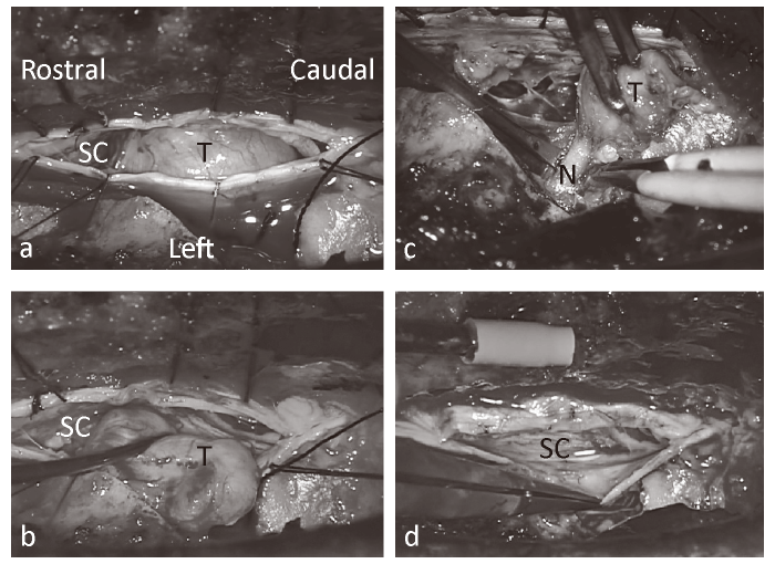
Fig. 3 Intraoperative microscope images. The spinal cord was strongly compressed by the tumor from the left side to the right and the ventral side. a, After opening the dura mater and arachnoid. b, Resection of tumor. c, Separation of outlet nerve root. d, Completion of tumor resection. SC, spinal cord; T, tumor; N, outlet nerve root.