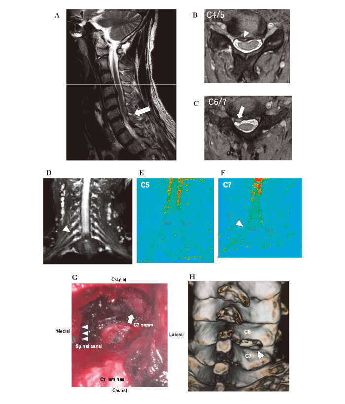
Fig. 8 Imaging and surgical findings of an example case. (A) Preoperative MRI sagittal image(T2-weighted image) of the cervical spine. Herniation of the C6-7 intervertebral disc can be observed(arrow) .(B) Preoperative axial MRI image of C4-5. Right-sided intervertebral disc herniation with mild spinal cord compression(arrowhead) was observed.(C) Preoperative axial MRI image of C6-7. Right-sided intervertebral disc herniation(arrowhead) was observed.(D) Cervical neurography(coronal image) . Swelling of the right C7 nerve(arrowhead) .(E) Cervical T2 mapping(coronal image) of C5 nerve There was no significant difference in T2 relaxation times(ms) between the C5 nerve roots on the left- and right sides(left: 89, right: 82) .(F) Cervical T2 mapping of C7 nerve. The T2 relaxation time was significantly prolonged in the right C7 nerve root(left: 127, right: 74) (arrowhead) .(G) Intraoperative photograph during microendoscopic surgery. We resected the inferior margin of the C6 lamina, and the superior margin of the C7 lamina, then removed the medial C6-7 facet joint, and exposed the area from the lateral margin of the dura mater(arrowhead) to the C7 nerve bifurcation(arrow) .(H) Postoperative 3D-CT image of the cervical spine. The fenestration of the inferior margin of the C6 lamina, the superior margin of the C7 lamina, and the medial third of the C6-7 facet joint(arrowhead) were confirmed. From Eguchi et al., modified with permission[18]
