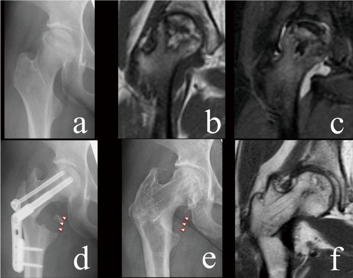
Fig. 2 Representative case of a 24-year-old woman with systemic lupus erythematosus who had osteonecrosis of the femoral head in her right hip. Preoperative X-ray (a) showed articular collapse within 2 mm (Stage 3A). MRI revealed a large necrotic area beyond the acetabular edge (Type C2) and bone marrow edema with joint effusion (b: T1 image, c: STIR image). A femoral curved varus osteotomy gained 32.4% of the intact ratio (the ratio of the non-osteonecrotic area to the weightbearing area) with 33o of varus correction (d). Fifteen years postoperatively, the femoral head had been preserved (e) with almost complete remodeling of the os calcar femorale (arrowhead). An MRI T1 image shows reduction of the area surrounding a low intensity band, indicating repair of the osteonecrosis (f).
