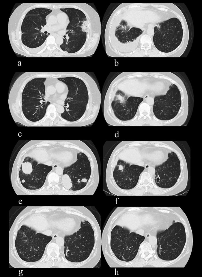
Fig. 4 Changes in chest CT images of Patient 2. (a) Axial image at the level of the arrowhead in Figure 3b. Right lung pleural effusion and left lung showing shadow. (b) Axial image at the level of the arrow in Figure 3b. Marked pleural effusion in the right lung. (c) Pleural effusion seen in (a), and (d) shadow seen in (b) have disappeared 9 months after high-dose steroid treatment. (e) Multiple nodules in both lungs at the time of the diagnosis of LPD. (f) The nodular shadows in both lungs were markedly reduced 5 months after discontinuation of MTX. The large para-aortic nodule in the left lung formed a cavity. (g) The nodular shadows had almost disappeared 11 months later. (h) LPD has not recurred during 51 months of follow-up.
