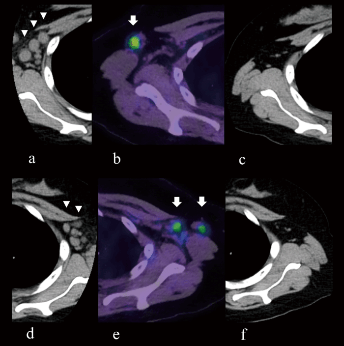
Fig. 6 Changes in CT and positron emission tomography (PET) / CT images of axillary lymph nodes in Patient 3. (a) Multiple enlarged lymph nodes in the right axilla (arrowheads) at the time of diagnosis of LPD. (b) Increased accumulation of fluorine 18-labeled fluorodeoxyglucose in the right axillary lymph node (arrow), but smaller size than 10 days prior. (c) The lymphadenopathy in the right lung has completely disappeared 7 months after discontinuation of MTX. (d) Multiple enlarged lymph nodes in the left axilla (arrowheads) at the time of diagnosis of LPD. (e) Increased accumulation of fluorine 18-labeled fluorodeoxyglucose in the left axillary lymph nodes (arrows), but smaller size than 10 days prior. (f) Lymphadenopathy in the left lung has completely disappeared 7 months after discontinuation of MTX.
