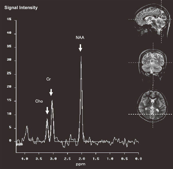
Fig. 1
Position of the volume of interest and example of a magnetic resonance sprectrum(MRS) of a 22-year-old normal man. MR images guided localization of the spectroscopic volume of interest in the occipital lobe are shown in transversal, coronal, and sagittal slices. The results of MRS are indicated as a spectrum of resonance(peaks) distributed along the X-axis labeled in parts per million(ppm). The amplitude of the resonance is measured on the y-axis using signal intensity in an arbitrary scale. The resonances of interest were N-acetylaspartate(NAA), creatine(Cr), and choline(Cho).