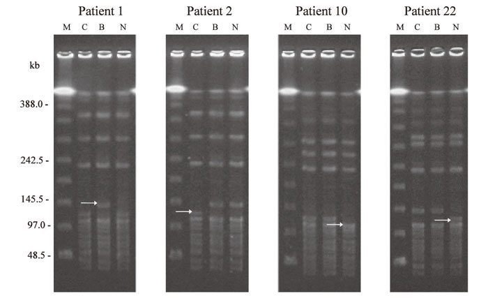
Fig. 3
Discrepancy of RFLP patterns of ApaI digested DNA between isolates from cerebrospinal fluid, blood, and nasopharynx of 4 patients. In all patients, a difference in the ~ 120 kb fragment was observed in 1 of 3 strains( arrow).
Patient 1, the isolate from the blood showed a singlebandshift of approximately +20 kb;
patient 2, the strain from the cerebrospinal fluid showed a shift of approximately -20 kb;
patients 10 and 22, strains from the nasopharynx showed a shift of approximately -20 kb shift.
Lanes:
C, isolate from cerebrospinal fluid;
B, from blood;
N, from nasopharynx;
M, lambda ladder molecular size marker.