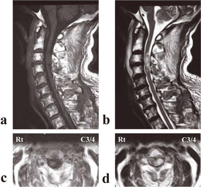
Fig.2
(a) T1-weighted midsagittal and (b) T2-weighted midsagittal of magnetic resonance imaging at the time of admission demonstrated a space-occupying lesion in the vertebral canal at the C3/4 level that showed an isointensity with the spinal cord on T1-weighted images and a high intensity on T2-weighted images. (c) T1-weighted axial and (d) T2-weighted axial views showed a round mass located on the left side of the spinal cord.