Fig. 1
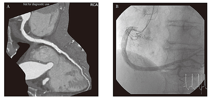
Typical Images Acquired with 320 Slice Computed Tomography (CT) and by Invasive Coronary Angiography (ICA)of a Patient with Chronic Atrial Fibrillation (CAF)(. Modified from Reference 39).
A: Typical Image of Coronary Arteries using Enhance 320 Slice CT.
Curved planar reformation image of the right coronary artery (RCA).
After acquiring plural heart beat data, the longest R-to-R interval data was manually selected and data were reconstructed by half reconstruction.
Even though this patient had CAF, there are no banding artifacts and image quality was excellent. There was no significant stenosis in any of the vessels on CT.
B: Images of ICA of the RCA, Acquired from the Same Patient as in Figure 1A.
There was also no significant stenosis found on ICA as seen on CT images shown in Figure 1A.