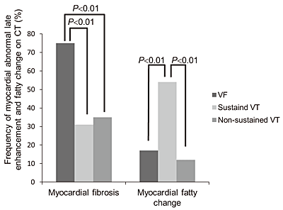
Fig. 7
Comparison of Frequency of Myocardial Abnormal Late Enhancement and Fatty Change on 320 Slice Computed Tomography (CT) among The Three“ Non Ischemic” Arrhythmia Groups (Ventricular Fibrillation (VF), Sustained and Non Sustained Ventricular Tachycardia (VT)) (Modified from Reference 35).
The frequency of myocardial abnormal late enhancement on CT was significantly higher in the non ischemic VF group (75%, all myocardial abnormal late enhancement was in the left ventricular myocardium) than in the non ischemic sustained VT group (31%) and the non ischemic non sustained VT group (35%) (both P<0.01). The frequency of myocardial fatty change on CT was significantly higher in the non ischemic sustained VT group (54%) than in the non ischemic VF group (17%) and the non ischemic non sustained VT group (12%) (both P<0.01).