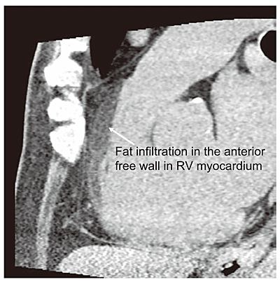
Fig. 8
Typical Non Enhanced Computed Tomographic (CT) Image of Fat Infiltration in the Anterior Free wall in the Right Ventricle (RV) Myocardium (Modified from Reference 36)
Left sagittal image.
A low CT area can be seen in the anterior free wall in RV myocardium.
The average CT value was -49HU.