


Chiba Medical J. 100E:1-6, 2024
doi:10.20776/S03035476-100E-1-P1
〔 Original Article 〕
Tatsuki Kobayashi1), Hiroaki Tsuruoka1), Kinatsu Fukushima2)
Atsushi Urata2), Masayuki Someya1), and Naohisa Kikuchi3)
1) Pediatric Orthopedics, Chiba Rehabilitation Center, Chiba 266-0005.
2) Prosthesis manufacturing facility, Chiba Rehabilitation Center, Chiba 266-0005.
3) Department of Rehabilitation, Chiba Rehabilitation Center, Chiba 266-0005.
(Received May 3, 2023, Accepted October 4, 2023, Published February 10, 2024.)
【Introduction】Pes planovalgus (PP) is the most common orthopedic complication of Down syndrome (DS). The frequency of PP in DS ranges from 19.9–51.4%, but there is no consensus on follow-up or therapeutic intervention. In this study, we investigated PP associated with DS, its symptoms, and the effects of orthotic therapy.
【Methods】The subjects were 52 children with DS (2–19 years old, mean 7.7 years old) who visited our department from July 2021 to June 2022. The parameters examined were the presence or absence of PP with visual inspection of the too-many-toes sign, symptoms such as foot pain, the presence or absence of orthotic therapy, and the presence or absence of arch formation. DS patients were followed-up annually. If the child had flat feet when they commenced walking, the family was instructed to create an arch support with felt on the insole of the shoe and to provide high-cut shoes. Patients would receive insoles made by the physician if they developed callouses, ingrown toenails, and/or hallux valgus, or if there was no change or worsening of their flat feet after reaching school age.
【Results】Observations of note were PP in 48 patients (92.3%), hallux valgus in 1 patient (2.9%), callouses in 2 patients (5.7%), ingrown toenails in 4 patients (11.4%), and foot pain in 2 patients (5.7%). Orthotic therapy was performed in 45 of 48 patients (93.8%). Arch formation was observed in 9 patients (18.8%), which included 8 of the 45 patients (mean age 7.8 years) who underwent treatment. Arch formation was confirmed in children who were 5–10 years old (mean 7.1 years old). There was no significant difference in arch formation between the treated and untreated groups (p=0.5).
【Conclusion】Many patients with DS have foot symptoms. Aggressive orthotic therapy for PP with DS may be useful.
pes planovalgus, Down syndrome, brace therapy
Pes planovalgus (PP) is the most common orthopedic complication of Down syndrome (DS), and the frequency of PP ranges from 19.9–51.4%[1]. It has been previously reported that PP associated with DS do not require aggressive treatment. However, Yamamoto et al. reported that approximately 70% of untreated cases did not form arches[2]. It may be useful to wear an orthotic early during the onset of walking. The purpose of this study was to investigate PP associated with DS, its symptoms, and the effects of orthotic therapy.
Participants
The study protocol was approved by the ethical review committee of Chiba Rehabilitation Center (July 29, 2022).
The subjects were 52 children with DS (2–19 years old, mean 7.7 years old) who visited our department from July 2021 to June 2022.
A flow diagram of the course of treatment is shown in Fig. 1. Patients were followed-up annually. If there was no arch when walking commenced, the family was instructed to create an arch support on the insole of the shoe using felt and the use of high-cut shoes was recommended. An insole was made by physicians for preschoolers when callouses, ingrown toenails, or hallux valgus was observed and for school age patients when there were no change or worsening of their PP.
The step-by-step construction of a felt arch is described below and depicted (Fig. 2).
① Measurement: the feet are placed on a piece of paper and the footprint is outlined using a pen. Landmarks of importance include the proximal head of the first metatarsal (A), the inferior aspect of the navicular (B), and the distal calcaneal bone (C). We show the exact locations on sole of the foot as A’, B’, and C’, respectively.
② Pattern making: D’, or the width of the felt arch, was approximately half the width of the foot at B’. A gentle curve was used to connect A’→D’ and D’→C’ and a straight line was used to connect A’→C’.
③ Material cutting: the shape of the arch support is cut out. A’→B’ should be twice as long as B’→C’.
④ The felt is scraped with a cutter: the felt should be contoured such that B’ represents the highest point.
⑤ Completion of insole (navicular pad) : the surface of the support should be made smooth overall.
⑥ Temporary alignment: shoes with heels firmly aligned should be used when inserting the felt insoles so that B’ is directly below B.
⑦ Determine the position: The shoes should be removed while the felt is held in place with one’s fingers to prevent it from moving while the position for its placement is marked.
⑧ Paste: The adhesive surface of the felt arch is exposed.
⑨ Completion: the arch is secured in place.
Data collection and analysis
The items examined were the presence or absence of PP by visual inspection of the too-many-toes sign[3]; the presence or absence of hallux valgus, callosities, ingrown toenails, and foot pain as foot symptoms; the presence or absence of orthotic therapy; and the presence or absence of arch formation. Differences between groups were tested using the t test and the chi square test for statistical analysis. P<0.05 was considered significant.
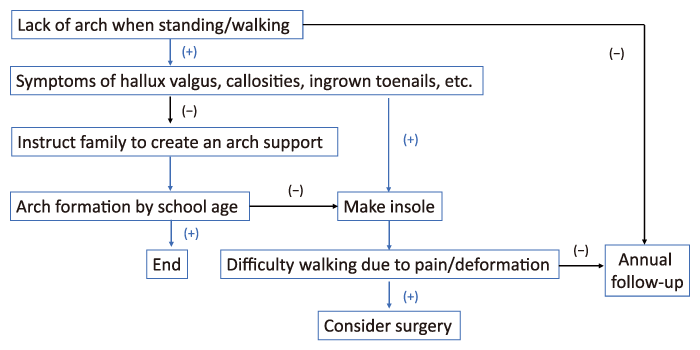
Fig. 1 Flow diagram of the course of treatment
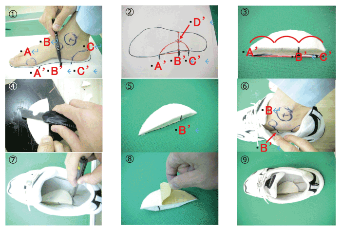
Fig. 2 How to construct a felt arch
Of the 52 patients who visited our department, 35 (67.3%) had PP at the time of examination, and 13 (25%) already had a history of PP, for a total of 48 patients (92.3%) with PP (Fig. 3). Among the patients with PP upon examination, hallux valgus was observed in 1 (2.9%), callosity was observed in 2 (5.7%), ingrown toenails were present in 4 (11.4%), and foot pain was reported for 2 patients (5.7%) (Table 1). One child had both ingrown toenails and foot pain. All children with foot symptoms at the time of examination had PP, with a prevalence of 7 of 35 (20%).
Of the 48 patients with PP at any time, 19 (39.6%) underwent treatment and 26 (54.2%) had received treatment in the past, for a total of 45 patients (93.8%) who were treated (Fig. 4). Three children did not receive orthotic therapy due to their parents’ wishes. The treatment was performed for all children with foot symptoms.
Arch formation was observed in 9 of the total 48 patients (18.8%, mean age 7.3 years), and no arch formation was evident in 43 patients (83%, mean age 7.7 years) (Fig. 5). There was no significant difference in age between those children with or without arch formation (p=0.67).
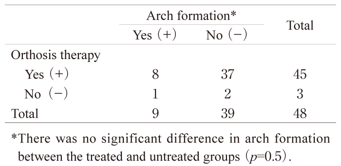
The relationship between orthotic therapy and arch formation has been summarized (Table 2). The mean age of the 8 patients who underwent orthotic therapy and formed foot arches was 7.8 years. Arch formation could be confirmed in children 5–10 years old (mean 7.1 years old). There was no significant difference in arch formation between the treated and untreated groups (p=0.5).
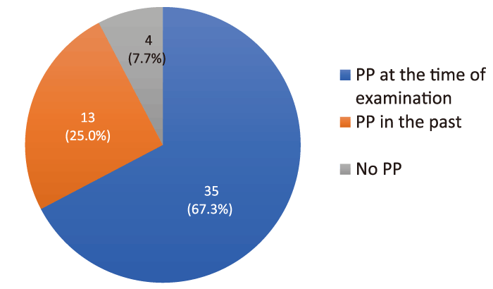
Fig. 3 Pes planovalgus
PP=pes planovalgus
Table 1 Occurrence of foot symptoms
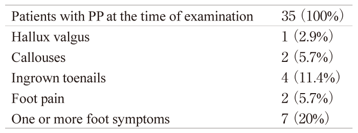
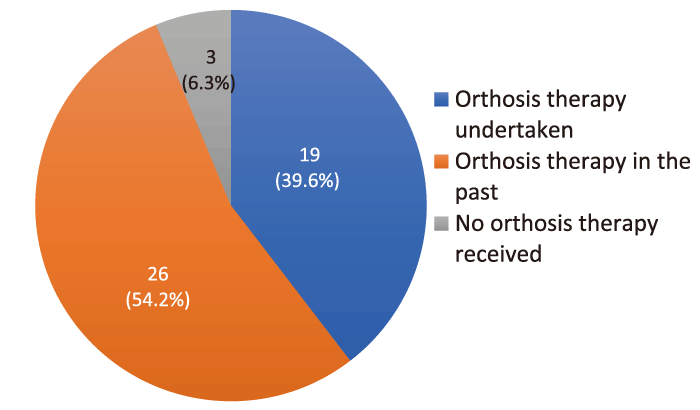
Fig. 4 Orthotic therapy
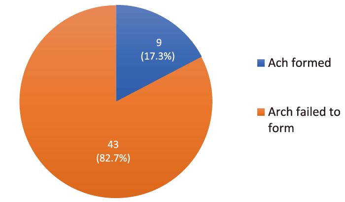
Fig. 5 Arch formation
Table 2 The relationship between orthosis therapy and arch formation
Yoshihide and colleagues have suggested that excessive radiation exposure and high costs associated with repeated follow-up examinations for PP should ideally be avoided[4]. Charlene et al. advocate for children with DS and recommend they undergo an annual musculoskeletal assessment as part of their health surveillance program[5]. During our study, patients were followed up annually for the presence of flat feet using a visual examination of the too-many-toes sign[3].
Of the 35 individuals with PP at presentation, 7 (20%) had foot symptoms. PP was observed in all 7 patients, and they all made orthotics. Previous reported studies indicated an association between flat feet and general joint laxity in the DS population[6-8].
Mitsuhiro et al. reported that the arch height index (AHI) and muscle mass of the soleus and tibialis posterior muscles decreased, while muscle mass of the flexor digitorum longus muscle increased in children with DS[9]. They also indicated that decreased AHI is associated with increased muscle mass of the flexor digitorum longus muscle in children[9].
Jung et al. showed shorter one-legged- and closed leg-stance times, in addition to shorter functional reaching, in children with DS than in children with typical development[10]. Jung and colleagues also demonstrated decreased walking speed in children with DS compared with children with typical development[10]. Walking speed was also associated with decreased plantar flexor muscle strength[11]. Previously, in children with DS and the complication of ectropion, the rate of PP was approximately 19.9–51.4%[1]. However, in a recent survey of 503 patients with DS, the prevalence of PP reached 91%, which is similar to the results of our study[5].
In the past, reports have indicated that minor orthopaedic problems are often underestimated and neglected despite the frequency of DS and severe associated pathologies[6]. It has also been reported that it would be better to have orthotic treatment than not to have orthotic treatment or that no treatment may suffice[12]. However, recently, there are reports that PP is common in children with DS; therefore, early consideration of orthotics and lifelong appropriate supportive footwear should be considered[5], and that a prescription for foot orthotics represents an early, evidence-based approach to slow down the onset of biomechanical abnormalities and potentially prevent related symptoms[13]. Previous studies have suggested that an insole can significantly decrease the eversion angle at the ankle joint[14] to improve the stability of the knee joint[15]. Not only can a foot arch lessen muscle fatigue, but it can also reduce energy consumption[16].
Yoshihide et al. recommended that subjects with DS should use an insole, even when a longitudinal arch fails to form despite the use of an orthotic; moreover, an insole prescription should continue to be renewed throughout life in order to maintain ambulatory ability[17].
We consider that orthotics are useful for PP associated with DS. However, when foot orthotics are made before a child reaches school age, they should be remade regularly as the child grows, which can be timeconsuming and costly. Therefore, we advise families to create felt arch supports for the insoles of shoes for preschool-aged patients with PP even when foot symptoms are absent.
We investigated PP associated with DS, its symptoms, and the effects of orthotics. PP was observed in more than 90% of patients. One-fifth of patients who had PP at the time of examination had foot symptoms. Treatment intervention was provided to 45 of 48 patients, and arch formation was observed in 9 of 48 patients (18.8%). Aggressive orthotic therapy for PP with DS may be useful.
TK and HT contributed to project development, data collection, and manuscript writing. KF, AU, and MS contributed to data collection. NK contributed as the scientific guarantor of this manuscript. All authors contributed to and approved the final manuscript.
No specific funding was used for this study.
The authors have no conflicts of interest.
The study protocol was approved by the ethics review committee of Chiba Rehabilitation Center. The participants were informed by opt out. This is not animal research.
All data generated or analyzed during this study are included in this published article.
Address correspondence to Dr. Tatsuki Kobayashi.
Pediatric Orthopedics, Chiba Rehabilitation Center, 1-45-2
Hondacho, Midori Ward, Chiba City, Chiba Prefecture
266-0005, Japan.
Phone: +81-43-291-1831.
Fax: +81-43-291-1857.
E-mail: tatsuki.chocolat@icloud.com
