


Chiba Medical J. 93E:1~9,2017
doi:10.20776/S03035476-93E-1-P1
[ Original Paper ]
Takashi Kaiho1), Shinji Yanagisawa1), Kazuyasu Shinmura1)
Ryo Okamoto1), Masaki Nishimura1), Souichi Kobayashi1)
Akira Okaniwa1), Shunichi Tsuchiya1), Kikuo Nagaoka2)
and Rikako Kobayashi2)
1) Department of Surgery, Kimitsu Chuo Hospital, Chiba 292-8535.
2) Nagaoka Clinic, Tokyo 136-0074.
(Received May 10, 2016, Accepted August 8, 2016)
Many bile duct cancers are diagnosed at the icteric stage. However, with recent advances in medical modalities, patients are being diagnosed and treated at the non-icteric stage, which leads to better prognosis. In the past 10 years, we performed resections in 44 cases of distal bile duct carcinoma: 11 in non-icteric and 33 in icteric patients. We retrospectively examined and compared the clinicopathological features of icteric and non-icteric patients. The first symptoms of non-icteric patients were abdominal pain and pyrexia. In regard to hepatobiliary enzymes, increased gammaglutamyltransferase(GGT) levels appeared more frequently than the other enzymes evaluated. For the non-icteric and icteric groups, the overall survival rates 1, 3 and 5 years after surgery were 90.9% and 72.7%; 90.9% and 42.5%; and 75.8% and 27.1%, respectively. The overall survival rate in non-icteric patients was significantly better than in icteric patients(P<0.05). Increased GGT level in patients without chronic hepatobiliary disease is an important component in the diagnosis of distal bile duct carcinoma at a non-icteric stage.
distal bile duct carcinoma, non-icteric bile duct carcinoma, gamma-glutamyltransferase, early bile duct carcinoma
Although many bile duct cancers are diagnosed at the icteric stage, with recent advances in medical modalities, such as multidetector-row computed tomography[1], peroral cholangioscopy[2], and intraductal ultrasonography[3], more cases are being diagnosed at a non-icteric stage. Several case reports have been published[4-6], but few involve non-icteric bile duct cancer cases obtained from a single institution [7.8]. Because icterus and obstructive jaundice are known to develop with disease progression, prognosis is likely to improve with the diagnosis of patients at a non-icteric stage. Here, we examined and compared the clinicopathological features of icteric and non-icteric distal bile duct carcinoma patients at our institution over the past 10 years.
From April 2003 to March 2013, we performed resections in 44 cases of distal bile duct carcinoma in Kimitsu Chuo Hospital: 11 cases were detected at a nonicteric stage(serum total bilirubin level < 1.0 mg/dl) at the first visit to the medical institution, and 33 cases were detected at the icteric stage(serum total bilirubin >1.0 mg/dl). Distal bile duct carcinoma predominantly occurs at the lower half of the extra-hepatic bile duct, with most lesions situated below the confluence of the cystic duct. Japanese society of Hepato-Biliary-Pancreatic Surgery defined distal bile duct carcinoma as occurring below the confluence of the cystic duct[9]. In this study, according to the American Joint Committee on Cancer(AJCC) cancer staging manual, the middle portion of the bile duct carcinomas were classified as their treatment, that is, a case with pancreaticoduodenectomy was treated as a distal one and a case with combined hepatic and hilar resection as a perihilar one[10].
We retrospectively compared patient characteristics, such as, age, sex, tumor maker level(carcinoembryonic antigen[CEA] and carbohydrate antigen 19-9[CA19-9]), and operative methods. Chief complaints and laboratory data at the first visit to the medical institution were also examined. Final pathological reports were reviewed to confirm the diagnosis of distal bile duct carcinoma. The pathological findings, such as macroscopic tumor type, histological tumor type, degree of bile duct invasion, regional lymph node metastasis, pathological stage and operative curability were investigated. We compared the operative morbidity and mortality, including postoperative pancreatic fistula(PF), delayed gastric emptying(DGE) and postoperative hepatic failure(HF). The definition of PF was by the international study group of postoperative pancreatic fistula(ISGPF)[11], DGE by the international study group of pancreatic surgery(ISGPS)[12] and HF by the international study group of liver surgery(ISGLS)[13]. We also surveyed the patient’s overall survival rate according to operative curability, pathological stages, and the presence or absence of icterus at the first visit. The extent of the disease, including the classification of lymph node metastasis and pathological stages, was defined according to the 7th edition of the Union for International Cancer Control(UICC), ICD-O C24.0. Operative curability was determined according to the currently used 6th edition of Japanese General Rules for Clinical and Pathological Studies on Cancer of the Biliary Tract[9].
Preoperative biliary drainage, endoscopically or percutaneously, was done in every cases except one in non-icteric group. In the non-icteric group, preoperative cholangiography revealed stenosis of the bile duct in some degree, so we drained all cases but one for prophylaxis of cholangitis.
The reginal lymph nodes removal was done in all of the resected cases around the hepatoduodenal ligament, the common hepatic artery and head of the pancreas, and more, around the superior mesenteric artery in the cases of pancreaticoduodenectomy.
The survival rate was calculated by the Kaplan-Meier method, and statistical analysis was performed using the log-rank test. Chi-square or Fischer's exact tests were used to compare qualitative parameters. P values of less than 0.05 were considered statistically significant.
The median observation periods for the non-icteric and icteric groups were 56.4 months(0.9~117.7) and 23.9 months(0.8~132.1), respectively. Although there were more male than female patients(27 : 6) in the icteric group, the patient gender ratio in non-icteric group was almost equivalent(6 : 5)(P=0.108). The patients' ages were comparable in both groups. The median level of tumor markers(CEA, CA19-9) was within the normal range in the non-icteric group, but CA19-9 was slightly elevated in the icteric group.
We resected tumors by using pylorus-preserving pancreaticoduodenectomy (PpPD) in 34 patients, Whipple's pancreaticoduodenectomy(PD) in 6 patients, duodenum preserving pancreas head resection(DPPHR)in a patient in non-icteric group, and extra-hepatic bile duct resection in three patients in icteric group.
DPPHR was done in a case of non-icteric group. She showed papillary tumor in the lower extrahepatic bile duct in preoperative cholangiography, and preoperative diagnosis was suspicion of carcinoma. Postoperative histological examination showed tumor was within mucosal layer with no lymph nodal metastasis, and histological curative resection(R0) was done. We resected bile duct only in three cases with the middle portion of extrahepatic bile duct carcinoma, because of age and poor general condition.
All patients underwent dissection of the regional lymph nodes. The portal vein was resected en bloc in two patients in the icteric group owing to direct carcinoma invasion(Table1). Six patients in the icteric group received postoperative adjuvant chemotherapy, four with Tegafur/Gimeracil/Oteracil(S-1) and two with gemcitabine. Each two patients in the both group underwent post-operative irradiation owing to positive margins at the hepatic duct.
Table. 1
Patients’ characteristics
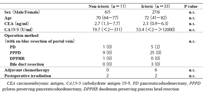
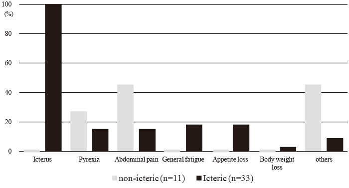
Fig. 1 The first symptoms in patients with the distal bile duct carcinoma
All of the icteric bile duct carcinoma patients first visited the medical clinic for icterus. Otherwise, in non-icteric patients, they visited the clinic for abdominal pain in five cases, pyrexia in three cases and three patients presented no clinical symptoms.
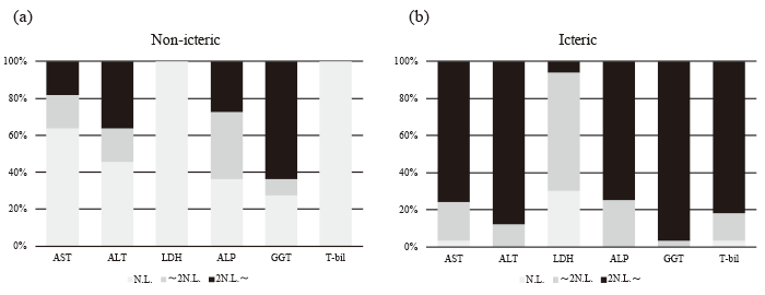
Fig. 2 Hepatobiliary enzyme profiles of the distal bile duct patients at the first visit to the medical institute
(a) In non-icteric patients, GGT increased more frequently than other enzymes, such as AST, ALT, LDH and ALP. (b) In icteric patients, the majority of the hepatobiliary enzymes showed abnormal levels, including serum total bilirubin.
GGT gamma-glutamyltransferase, AST aspartate aminotransferase, ALT alanine aminotransferase, LDH lactate dehydrogenase, ALP alkaline phosphatase.
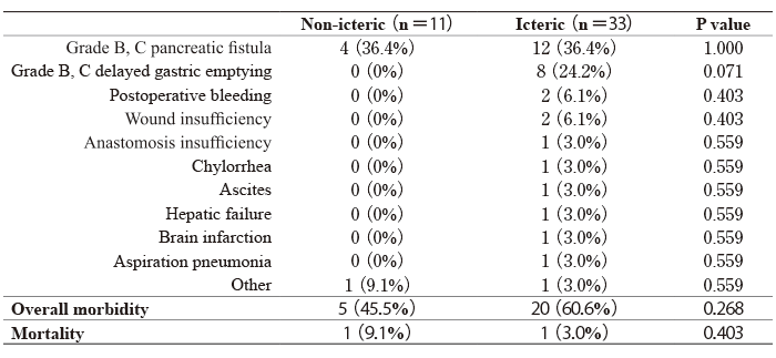
One hospital death occurred in each group. One patient in the icteric group died 25 days after PpPD because of hepatic failure. He developed preoperative liver cirrhosis derived owing to hepatitis C virus infection, and postoperative uncontrollable ascites occurred. Another patient in the non-icteric group died 26 days after PpPD because of aspiration pneumonia. He aspirated vomitus, which induced cardiopulmonary arrest on the 5th postoperative day. Grade B or C pancreatic fistula[11] occurred in 12 cases(36.4%)in the icteric group, and in 4 cases(36.4%) in the nonicteric group, respectively; grade B or C delayed gastric emptying[12]occurred in 8 cases(24.2%) in the icteric group and in none of the cases in the non-icteric group. Other postoperative complications are listed in Table 2. Overall morbidity was 60.6% in the icteric group and 45.5% in the non-icteric group(n.s.). The median hospital stay was 33(range 13-113) and 35(16-139) days in the non-icteric and icteric groups, respectively.
Table. 2
Postoperative morbidity and mortality
Average tumor diameter in the non-icteric group was 28.0±15.0mm, and 33.6±18.5mm in the icteric group(n.s.). With regards to macroscopic tumor type, papillary-expanding, papillary-infiltrating and nodularexpanding types accounted for 54.5% of the tumors in the non-icteric group; nodular-infiltrating and flatinfiltrating types accounted for 84.8% of the tumors in the icteric group. Regarding the histological tumor type, papillary adenocarcinoma(pap) and well-differentiated tubular adenocarcinoma(tub1) accounted for 90.9% of the tumors in the non-icteric group; poorly differentiated adenocarcinoma(por) and moderately differentiated tubular adenocarcinoma(tub2) accounted for 66.6% of the tumors in the icteric group. One patient in the icteric group had an adenosquamous carcinoma of the bile duct. The depth of bile duct wall invasion at the level of T1 or T2 accounted for 90.9% of the cases in the non-icteric group; Moreover, 72.7% of the cases in icteric group were over T3, and 81.8% of the nonicteric patients had no regional lymph node metastasis. In contrast, 54.5% of the icteric bile duct carcinoma patients exhibited metastasis to regional lymph nodes.
In terms of pathological stage, 72.7% were under Stage IB in the non-icteric group, whereas 78.8% were over Stage IIA in the icteric group(Table 3).
Table. 3
Pathological findings
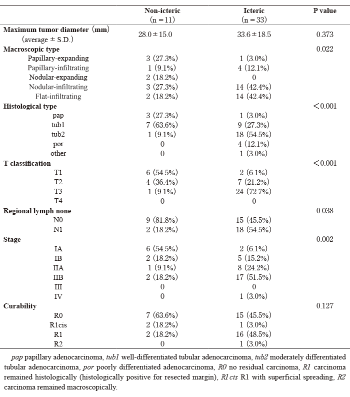
With regards to overall survival, Stage IA patients had significantly better prognoses than Stage IIA (P<0.05) and IIB (P<0.01) patients (Fig. 3).
Histologically curative surgery(R0) was performed in 63.6% and 45.5% of the patients in the non-icteric and icteric groups, respectively(Table 3). The R0 group showed significantly better prognosis than the non-curative group(R1, R1cis, R2)(P<0.01)(Fig. 4). The overall survival rates 1, 3, and 5 years after surgery were 90.9% and 72.7%, 90.9% and 42.5%, and 75.8% and 27.1% in the non-icteric and icteric groups, respectively. The overall survival rate in patients who underwent resection for non-icteric distal bile duct carcinoma was significantly better than that in icteric patients(P<0.05)(Fig. 5).
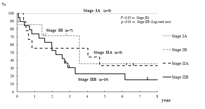
Fig. 3 Overall survival according to the pathological stage
Stage IA patients exhibited significantly better prognosis than Stage IIA (P<0.05) and Stage IIB (P<0.01)patients.
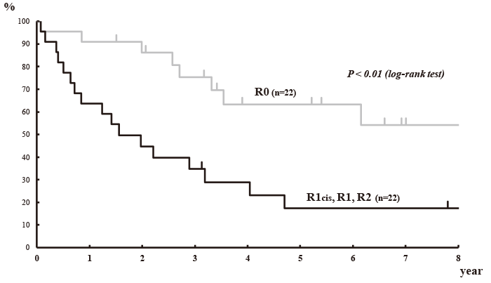
Fig. 4 Overall survival according to the curability
Patients in the histologically curative operation(R0)group(grey line) showed significantly better prognosis than those in the non-curative group(R1, R1cis, R2)(black line; P<0.01). <
R0 no residual carcinoma, R1 carcinoma remained histologically(histologically positive for resected margin), R1 cis R1 with superficial spreading, R2 carcinoma remained macroscopically.
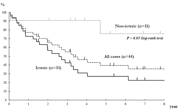
Fig. 5 Overall survival of the distal bile duct carcinoma patients between non-icteric and icteric patients
Overall survival rates 1, 3 and 5 years after surgery were 90.9% and 72.7%; 90.9% and 42.5%; and 75.8% and 27.1% in the non-icteric and icteric groups, respectively.
The overall survival rate of the resected patients with non-icteric distal bile duct carcinoma(grey line) was significantly better than that of icteric patients(black line; P<0.05).
Early gastric or colorectal cancers are defined as lesions in which the depth of wall invasion is within the submucosal layer, regardless of lymph node metastasis[14,15]. These early carcinomas show comparably better prognoses than those that are more advanced.Screening gastro fiberscope or fecal occult blood tests have contributed to improved detection of these carcinomas[16,17] On the other hand, there is no clear definition of early bile duct carcinoma, although some authors advocate defining“ the early bile duct carcinoma” as a lesion in which the depth of wall invasion is within the fibro-muscular layer, and report its favorable prognosis[3,18-20].
Most bile duct carcinoma patients visit medical institutions for icterus. Distal bile duct carcinoma often requires PD or PpPD for curative resection-operative procedures that have high morbidity and mortality [21,22]. Even if a clinically curative operation is performed, it is difficult to obtain histological negative margins [23].
Icterus inevitably appears in distal bile duct carcinoma with progression of the disease, whereas in perihilar bile duct carcinoma, icterus does not appear until the tumor invades the confluence. Consequently, icterus may appear somewhat earlier in distal bile duct carcinoma than in perihilar carcinoma. If distal bile duct carcinoma is diagnosed earlier, at a non-icteric stage, the prognosis is better. As the levels of two tumor markers, CEA and CA19-9, were within the normal range in the majority of the non-icteric bile duct patients evaluated, they contributed little to a meaningful diagnosis.
GGT is a membrane-bound enzyme that exists in several tissues, such as the kidney, pancreas, and liver, and plays a role in the metabolism of glutathione and in the facilitation of amino acid transport. It is known that serum GGT levels increase in hepatobiliary diseaseespecially alcoholic liver disease and cholestasisbut not in renal disease. GGT is synthetized in the endoplasmic reticulum of hepatocytes and is then released and localized in the membranes of the bile canaliculi in the liver, or in the epithelium of the bileduct[24]. In Korean report, among the medical records of 2024 subjects who attended the institution for medical check-up, 78 subjects were a positive for hepatitis C virus antibody and/or hepatitis B virus surface antigen and/or liver cirrhosis, 579 subjects were a daily alcohol intake of 20g or more. In the rest of 1367 subjects, 352 subjects(25.7%) showed abnormal hepatic enzyme/ GGT, 10 subjects were with incomplete data. Among the rest of 1005 subjects, serum alanine aminotransferase (ALT) and GGT concentration within the reference ranges correlated with the incidence of nonalcoholic fatty liver disease(NAFLD) and metabolic syndrome in a dose-dependent manner[25].
Serum bilirubin levels increase in obstructive jaundice, but not in the case of partial obstruction of the bile duct. Thus, in the case of perihilar bile duct carcinoma, obstruction of unilateral hepatic ducts, i.e., either the right or left hepatic duct, does not lead to increased serum bilirubin levels until the tumor has invaded the confluence. Sugiyama et al. have reported cases of extrahepatic bile duct carcinoma without jaundice, but most of these lesions(89%) were in the upper third of the extrahepatic bile duct[7]. Ito et al. also reported clinicopathological features of extrahepatic cholangiocarcinoma without jaundice, and 40% of these were perihilar lesions[8]. Thus, in these reports, carcinomas that were not associated with jaundice were mainly located in the upper bile duct.
On the other hand, ALP or GGT levels increase even in the case of hemi obstruction. In this study, GGT increased more frequently than the other hepatobiliary enzymes evaluated in the patients without jaundice. GGT is thought to be the most sensitive hepatocytereleased enzyme marker of bile duct compression.
In this study, non-icteric bile duct carcinoma was characterized by:(1) macroscopic tumor types consisting of papillary-expanding, papillary-infiltrating, and nodular-expanding tumors(54.5%)(; 2) histological tumor types comprising papillary adenocarcinoma and well-differentiated tubular enocarcinoma(90.9%); (3) depth of bile duct wall invasion at level T1 or T2(90.9%);(4) no regional lymph node metastasis (81.8% of patients); and(5) pathological stages IA and IB(72.7% of the cases), indicating that non-icteric patients do not always have early-stage tumors. On the contrary, the icteric bile duct carcinoma cases in this study were characterized by:(1) macroscopic tumor types consisting of nodular-infiltrating type and flatinfiltrating type tumors(84.8%);(2) histological tumor types comprising poorly differentiated adenocarcinoma and moderately differentiated tubular adenocarcinoma (66.6%);(3) depth of bile duct wall invasion over T3 (72.7%)(; 4) regional lymph node metastasis(54.5% of patients); and(5) pathological stage greater than Stage II disease(78.8% of patients). Although Sugiyama et al. and Ito et al. reported that there were no significant differences in the pathological findings between nonicteric and icteric patients[7,8], there were obvious differences between these patient groups in our study. A reason for this may be that almost half of the cases(40-89%) in the previous studies were proximal bile duct carcinomas, making it possible that more advanced cases were included in the non-icteric groups.
The R0 resection rate was 63.6% in the non-icteric group and 45.5% in the icteric group; the overall curative resection rate was 50.0%, which is comparable to previously reported cases(30.4%-68.1%)[21,23]. Even in the non-icteric group, the curative resection rate was somewhat low(63.6%), probably because of histologically positive margins at the surgical site in four patients(36.4%), two of whom(18.2%) showed superficial mucosal spreading of carcinoma. It is well known that the intraductal papillary growth of bile duct carcinoma is likely to spread widely and superficially [26]. The curative operation group showed significantly better prognosis than the non-curative group(P<0.01), emphasizing the importance of a negative margin during the surgical procedure.
The overall survival rate of resected patients with non-icteric distal bile duct carcinoma was significantly better than that of icteric patients(P<0.05). Sugiyama et al. also reported significantly higher survival rates in non-icteric patients compared with icteric patients(50% vs. 22% at 5 years)[7]. Relatively better prognosis in non-icteric bile duct carcinoma is thought to be related to early stage, well-differentiated carcinoma and less regional lymph node metastasis. Because of the soft pancreas, the rates of grade B and C postoperative pancreatic fistula were higher in this study compared to those reported in other cases, but were equivalent in both groups. The overall morbidity rate was lower in the nonicteric than icteric group(45.5% vs. 60.6%), but not significant. Two hospital deaths occurred in this study; this 4.5%(2/44) death rate is similar to that reported in the Japanese national web-based data of extrahepatic bile duct carcinoma[22]. In terms of prognosis and operative morbidity, it is critical that we diagnose and treat distal bile duct carcinoma patients at a non-icteric stage.
Therefore, here we emphasize the importance of the increase in GGT levels in patients without chronic liver disease, for example, those with chronic hepatitis, alcoholic liver disease, or nonalcoholic fatty liver disease, as a tool in diagnosing distal bile duct carcinoma at a non-icteric stage.
Address correspondence to Dr. Takashi Kaiho.
Department of Surgery, Kimitsu Chuo Hospital, 1010 Sakurai, Kisarazu, Chiba 292-8535, Japan.
Phone: +81-438-36-1071. Fax: +81-438-36-0890.
E-mail:kaihot@jcom.zaq.ne.jp
