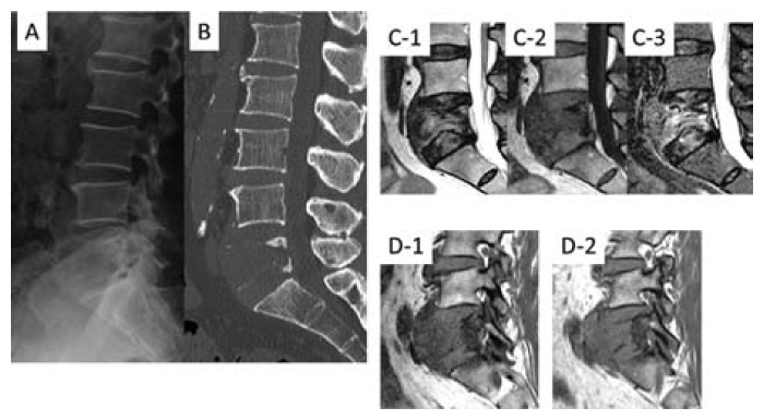
Fig. 2 Imaging findings at the time of admission
X-ray(A) and sagittal computed tomography(B) images demonstrate that most of the L5 vertebral body was destroyed. Sagittal T1-weighted(C-1), T2-weighted(C- 2), short tau inversion recovery sequence(C-3) magnetic resonance images demonstrate that the tumor had invaded most of the L5 vertebral body. Sagittal T1-weighted images of the bilateral intervertebral foramen(D-1; Right side, D-2; Left side) demonstrate that the tumor had severely compressed the L5 nerve roots, bilaterally.
