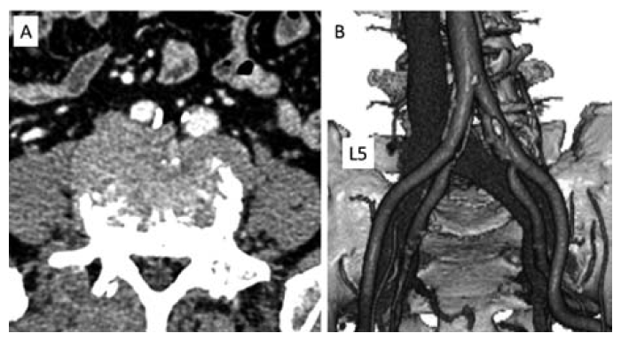
Fig. 3 Computed tomography angiography imaging studies
Axial(A) and 3-dimensional(B) images show the bifurcation of the descending aorta and inferior vena cava in front of the L5 vertebral body.
Fig. 3 Computed tomography angiography imaging studies
Axial(A) and 3-dimensional(B) images show the bifurcation of the descending aorta and inferior vena cava in front of the L5 vertebral body.