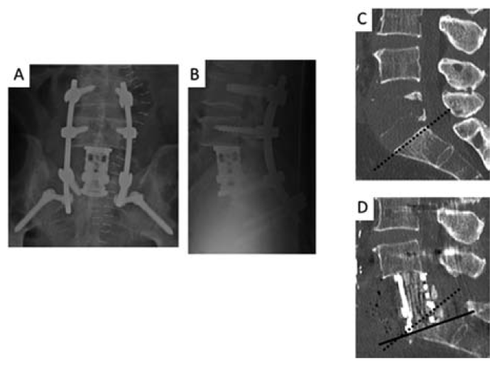
Fig. 4 Postoperative imaging studies
Anteroposterior(A) and lateral(B) x-ray images show the inserted expandable cage, a 16-degree lordotic cage, fixed in place. Preoperative(C) and postoperative(D) sagittal computed tomography images demonstrate the changes after 20-degrees partial wedge resection of the S1 head side endplate. The dotted line shows the angle of the S1 head side end plate before the operation. The black line indicates the angle of the endplate of the S1 head side after surgery.
