


Chiba Medical J. 93E:45~51,2017
doi:10.20776/S03035476-93E-5-P45
[ Original Paper ]
Junichi Nakamura1), Nobuyasu Ochiai1), Seiji Ohtori1),
Sumihisa Orita1), Shigeo Hagiwara1), Hironori Yamazaki1),
Takane Suzuki2), and Kazuhisa Takahashi1)
1) Department of Orthopaedic Surgery, Graduate School of Medicine, Chiba University, Chiba 260-8670.
2) Department of Bioenvironmental Medicine, Graduate School of Medicine, Chiba University, Chiba 260-8670.
(Received March 13, 2017, Accepted April 4, 2017)
Objectives: The aim was to evaluate the safety and efficacy of extracorporeal shock wave therapy (ESWT) for osteonecrosis of the femoral head.
Methods: This was a phase I clinical trial and prospective case-control study. Large osteonecrotic areas with advanced collapse were included. ESWT was applied from anterior to posterior with 5000 impulses. Energy flux density was started at level one(0.03mJ/mm2) and advanced to level seven(0.36 mJ/mm2). A historical cohort was matched to this treatment group.
Results: ESWT and natural history groups each consisted of 28 hips. There were no obvious complications such as progression of osteonecrosis or neurovascular injury. ESWT group showed a gradual improvement in hip score and pain score, and these scores were significantly different than those of the natural history group at the end of treatment(p=0.034 and 0.019, respectively). The survival rate for total hip arthroplasty at two years was not significantly different between ESWT group and the natural history group(35.1% versus 24.7%, p=0.749). However, Cox regression analysis revealed that the type C2 hip was an independent and significant prognostic factor with an 8.6- fold higher hazard ratio than type C1(p=0.004).
Conclusions: ESWT showed safe and effective for osteonecrosis of the femoral head.
osteonecrosis of the femoral head, extracorporeal shock wave therapy
Osteonecrosis of the femoral head(ONFH) causes joint destruction after articular collapse, followed by impairment of quality of life due to hip pain and gait disturbances in young and active individuals[1]. Corticosteroid therapy is an influential factor in the development of ONFH; the incidence of ONFH was reported as 41% in adult systemic lupus erythematosus (SLE) patients[2]. In general, conservative treatment has little effect on the symptoms and surgical treatment often is required. The cumulative surgical frequency has been reported as 67% at 5 years after articular collapse in ONFH[3]. Preservation of the joint using regenerative medicine or femoral osteotomy has been proposed, but there are several limitations to these approaches[4-8]. Total hip arthroplasty(THA) is a promising surgical option although implant loosening requires revision surgery[9-10]. On the other hand, not all patients with osteonecrosis undergo surgery because of high risks associated with generally poor health related to the underlying disease. Therefore, it is important to establish safe and effective conservative treatment for ONFH.
Extracorporeal shock wave therapy(ESWT) has been applied clinically to chronic painful musculoskeletal disorders[11-12]. ESWT induces degeneration of free nerve endings and sensory nerve fibers, followed by a decrease in the number of dorsal root ganglion neurons [13-15]. As hip pain is caused by invasion of free nerve endings and sensory nerve fibers to the synovium[16], we hypothesized that ESWT would provide pain relief from the collapsed femoral head in ONFH. The purpose of this study was to evaluate the safety and effectiveness of ESWT for ONFH.
The protocol for this phase I clinical trial and prospective case-control study was approved by the institutional review board and the participants gave written informed consent(University Hospital Medical Information Network, UMIN000020197). The primary end point was to confirm safety and the secondary end point was to determine effectiveness.
Patients with ONFH diagnosed by the classification of the Japanese Ministry of Health, Labor and Welfare (JMHLW)[17-18], with large necrotic areas of type C1 or C2, and with advanced collapse at stage 3B or 4 were included. It was reasonable ethically to study safety in these patients because they were, in principal, candidates for THA. In case of therapeutic failure, THA would be an acceptable salvage procedure. Exclusion criteria included patients with skeletal immaturity, previous or current infections, unfavorable skin conditions, or patients with hemorrhagic diathesis. The JMHWL classification was used to categorize lesion size and stage for articular collapse. A type A lesion occupies the medial one-third or less of the weight-bearing portion of the femoral head; a type B lesion occupies the medial two-thirds or less of the weight-bearing portion; a type C1 lesion occupies more than the medial two-thirds of the weight-bearing portion of the femoral head but does not extend laterally to the acetabular edge; and a type C2 lesion extends laterally to the acetabular edge. Stage 1 is defined by normal findings on radiograph, but specific findings on magnetic resonance imaging(MRI), a bone scan, or by histology; stage 2 is defined by demarcating sclerosis without collapse of the femoral head; stage 3A involves collapse of the femoral head less than 3mm in diameter; stage 3B involves collapse of the femoral head more than 3mm in diameter; and stage 4 is defined by osteoarthritic changes with joint space narrowing.
From August 2010 to September 2012, ESWT was performed after informed consent for all the participants in the shock wave group. A historical cohort of patients with ONFH from the same institution was matched to this treatment group by age, gender, etiology, and the type and stage classification of JMHLW. ESWT and natural history groups each consisted of 28 hips. Sensitivity analysis and power analysis could not be performed because there were no case-control studies in the previous literature. Julious[19] recommended a sample size of 12 per group for a pilot study when there is no prior information on which to base a sample size. The sample size of 28 per group was based on the number of new patients annually(20-30 patients/year) in our hospital. ESWT was administered with Dornier EposTM(Dornier, Germany), which generates a shock wave by electromagnetic induction. The junctional zone between the osteonecrotic and viable bone of the femoral head was identified radiologically using an image intensifier. One point within the junctional zone was chosen with a metallic pin under the image intensifier, and marked on the skin in the groin. The treatment was performed on a bed with the patient in the supine position in the outpatient clinic. Patients were awake without any anesthesia so that they could comment on whether the shock wave was reproducing their hip pain. The femoral artery was palpated and protected from direct contact with the shock wave tube during the course of treatment. Ultrasound jelly was used on the skin in contact with the shock wave tube. Shock waves were applied from anterior to posterior with 5000 impulses at a rate of 240 impulses/min with a 4×25mm focus area. Energy flux density was started at level one(0.03mJ/mm2) and advanced as tolerated to level seven(0.36 mJ/mm2). If pain from osteonecrosis was not reproduced by the shock wave, the shock wave tube was moved to find a point that reproduced the pain of osteonecrosis. ESWT was applied three times every one to two months. The follow-up period was two years.
Outcome measures were assessed by hip score using the Japanese Orthopaedic Association(JOA) [20], PainDETECT(PD)[21], Visual Analogue Scale (VAS), total energy of ESWT, and the progression of femoral head collapse. The Fisher exact probability test was calculated for gender difference, underlying disease (SLE versus non-SLE) and JMHLW classification using IBM SPSS Statistics 16.0(Chicago, Illinois). Pearson's χ2 test was done for etiology(Steroidal, alcoholic, or" idiopathic"). The Mann-Whitney U test was done for age, JOA hip score, PD score, and VAS. Friedman test was calculated for parameters at initial examination, the first, the second, and the third ESWT. The Kaplan-Meier method with log rank test and Cox regression analysis were calculated for the final end point of total hip arthroplasty. A p-value<0.05 was considered significant.
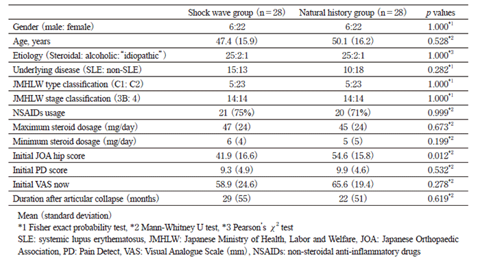
ESWT and natural history groups each consisted of 28 hips. Gender, age, etiology, underlying disease, classification of osteonecrosis, use of non-steroidal anti-inflammatory drugs, steroid dosage, duration after articular collapse, initial PD score and initial VAS were not significantly different between groups at the start of therapy(Table1). The initial JOA hip score was significantly worse in ESWT group than in the natural history group(41.9 points versus 54.6 points, p=0.012).
Patient characteristics
There were no obvious complications such as rapid progression towards collapse of the osteonecrotic lesion or neurovascular injury after ESWT at the two-year follow up.
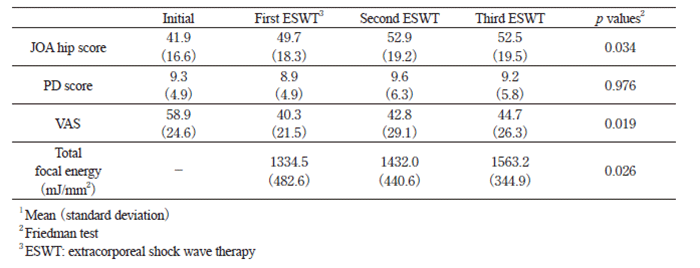
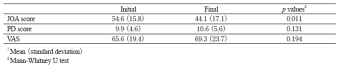
ESWT group showed a gradual improvement in JOA hip score and VAS, and these scores were significantly different than those of the natural history group at the end of treatment(p=0.034 and 0.019, respectively, Table2). The focal energy of the shock wave significantly increased over the course of the treatment(p=0.026). On the other hand, the JOA hip score deteriorated in the natural history group(p=0.011, Table3). Thus, the amount of change in the JOA hip score that occurred within ESWT and natural history groups was significantly different by the end of treatment(+9.1 points versus -10.5 points, p=0.001). The groups also differed significantly on the VAS score (-9.0 points versus +3.6 points, p=0.021)
Time course of shock wave group1
Outcome of the natural history group1
The survival rate for THA was not significantly different between ESWT group and the natural history group(p=0.749, Figure 1). However, the survival rate was significantly better in type C1 than in type C2 hips by the JMHLW classification(p=0.001, Figure 2). The survival rate also was significantly better in stage 3B than in stage 4 using this classification(p=0.027, Figure 3). Cox regression analysis revealed that the type C2 hip was an independent and significant prognostic factor for THA with an 8.6 fold higher hazard ratio than type C1(p=0.004)
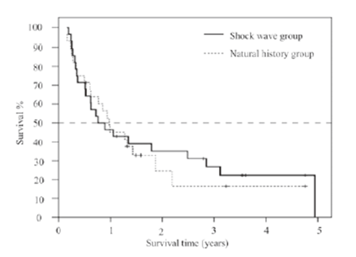
Fig. 1 Survivorship of shock wave and natural history groups
When the end point was total hip arthroplasty, survival at one year was 42.9% in the shock wave group and 45.1% in the natural history group; survival at two years was 35.1% in the shock wave group and 24.7% in the natural history group, without a significant difference(Kaplan- Meier and log rank test, p=0.749).
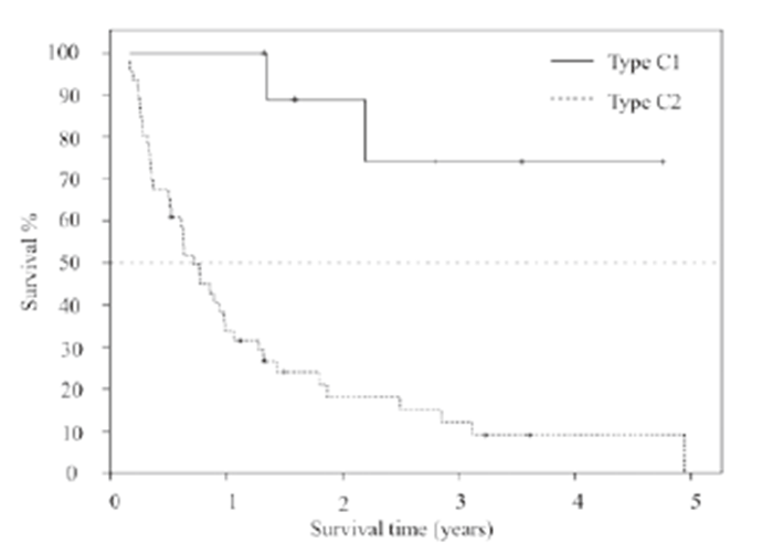
Fig. 2 Survivorship of type C1 and type C2 hips by JMHLW classification
When the end point was total hip arthroplasty, survival at one year was 100% in type C1 and 33.8% in type C2; survival at two years was 88.9% in type C1 and 18.0% in type C2, with a significant difference(Kaplan-Meier and log rank test, p=0.001).
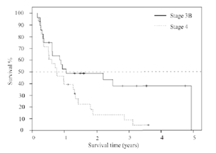
Fig. 3 Survivorship of stage 3B and stage 4 hips by JMHLW classification
When the end point was total hip arthroplasty, survival at one year was 52.5% in stage 3B and 39.3% in stage 4; survival at two years was 48.8% in stage 3B and 13.5% in stage 4, with a significant difference(Kaplan-Meier and log rank test, p=0.027).
ESWT demonstrated a safety profile without complications and improved the JOA hip score and VAS, indicating ESWT can be an optional conservative treatment for advanced ONFH. However, ESWT did not improve the natural history of the osteonecrosis nor prevent an eventual THA. ESWT was first applied for ONFH in 1998[22]. In Japan, clinical use of ESWT was approved in 2008. Experimental work has indicated some palliative effect for ESWT[13-15], although clinical trials are limited. Thus, the goal of our study was to evaluate its safety and effectiveness. ESWT offers several important advantages over conventional surgical treatment: it is a noninvasive treatment with a substantially reduced incidence of complications, and it does not make the technique of THA difficult, if it becomes necessary, because skeletal alignment has not been altered.
Ludwig et al.[22]reported favorable results with ESWT in which 14 of 21 patients improved in pain and hip score over one year, but one-third(7 patients) returned to their previous symptoms. Wang et al.[23] reported that 79% of the hips were improved, 10% were unchanged, and 10% were worse with ESWT in a prospective study. Wang's group performed ESWT under general anesthesia with 6000 impulses of 0.62 mJ/mm2 energy flux density(total dose: 3720 mJ/mm2) at one time. In Japan, the maximum energy flux density is limited by law to 0.36 mJ/mm2. In our study, the energy flux density began at 0.03mJ/mm2 and increased to 0.36 mJ/mm2 without anesthesia, using three treatments of 5000 pulses each. The difference in protocol may be one reason our results are not as positive as Wang's. Hausdorf et al.[24]showed that 49.2% of the shock wave pressure occurred 10 mm inside the femoral head, and the pressure increased with energy. Therefore highenergy shock waves may be more effective to regenerate bone.
Another factor may be the extent of osteonecrosis. Ludwig et al.[22]indicated ESWT was successful for the early stage osteonecrosis. Vulpiani et al.[25] reported patients at stages 1 or 2 achieved significantly better results than patients at stage 3 at all time points. At a minimum two-year follow up, 10 of the 15 stage 3 patients(67%) received arthroplasty, whereas stage 1 and 2 lesions were unchanged on radiographs and on magnetic resonance images. Wang et al.[26]reported total hip replacement after ESWT in 12% of patients with SLE at a minimum two-year follow up, but they did not include the patients at stage 4. Our study included only advanced or end stage osteonecrosis patients and found the prognosis at stage 4 to be poor. We believe this is another reason for our poor outcome.
Type C2 hips classified using the JMHLW classification are an independent prognostic indicator for THA. It is widely accepted that type C2 hips often result in articular collapse[27]and that after collapse the survival rate is 33% at 5 years and 22% at 10 years [3]. Kaplan-Meier curves of the shock wave and natural history groups were almost the same(Figure 1), indicating ESWT does not change the risk for a collapsed femoral head.
The mechanism of ESWT is of great interest. Wang et al.[28]documented that local ESWT application led to significant elevations of serum nitric oxide, angiogenic, osteogenic, and anti-inflammatory factors in patients with osteonecrosis of the femoral head. Wang et al.[29]also showed significantly more viable bone and less necrotic bone in the femoral head after ESWT by histopathological examination, and significant increases in cytokines for angiogenesis, such as von Willebrand factor and vascular endothelial growth factor, than in the controls. In the current study, osteogenesis was not observed after ESWT, but some pain relief was achieved.
There are several limitations to this study. First, because this was a phase I clinical trial and the primary end point was safety of ESWT, only advanced stage patients were included. Further study is needed to confirm its effectiveness for early stage osteonecrosis. Second, the optimal protocol for ESWT has not been established; the number of pulses, energy flux density, number of treatments, treatment interval, and effects of anesthesia are all variables that should be evaluated. A multi-center study to define the best treatment protocol will be essential. Third, sensitivity analysis and power analysis could not be done. Based on our results, sample size could be estimated for a future study. Fourth, a follow-up period of two years is short and the long-term outcome is still controversial. ESWT is not curative, but is palliative. However, patients with ONFH often have general complications and are at high risk for surgery. ESWT can be a safe treatment option for such patients.
In conclusion, ESWT improved pain and functional hip score in patients with ONFH without complications, indicating it can be an optional conservative treatment. However, the outcome of ESWT did not slow the progression towards THA. The type classification of the hip by JMHLW was an independent prognostic factor for THA.
This work was supported by a grant from Inohana Shogakukai and JSPS KAKENHI Grant Number 25870125.
Address correspondence to Dr. Junichi Nakamura.
Department of Orthopaedic Surgery, Graduate School of Medicine, Chiba University, 1-8-1, Inohana, Chuou-ku, Chiba 260-8670, Japan.
Phone: +81-43-226-2117. Fax: +81-43-226-2116.
E-mail:njonedr@chiba-u.jp
