


Chiba Medical J. 93E:53~57,2017
doi:10.20776/S03035476-93E-5-P53
[ Case Report ]
Takaki Inoue1), Sumihisa Orita1)*, Hiroto Kamoda2),
Yoshihiro Sakuma1), Kazuhide Inage1), Jun Sato1),
Kazuki Fujimoto1), Yasuhiro Shiga1), Hirohito Kanamoto1),
Koki Abe1), Kazuyo Yamauchi1), Junichi Nakamura1),
Yusuke Matsuura1),Yasuchika Aoki3), Yawara Eguchi4),
Kazuhisa Takahashi1), Shigeo Hagiwara1), Takeo Furuya1),
Masao Koda1), Masahiko Suzuki1) and Seiji Ohtori1)
1) Department of Orthopaedic Surgery, Graduate School of Medicine, Chiba University, Chiba 260-8670.
2) Department of Orthopaedic surgery, Chiba Cancer Center, Chiba 260-8717.
3) Department of Orthopaedic surgery, Eastern Chiba Medical Center, Togane 283-8686.
4) Department of Orthopaedic surgery, Shimoshizu National Hospital, Yotsukaido 284-0003.
(Received March 17, 2017, Accepted April 21, 2017)
A 75 year-old woman was referred to our clinic for a solitary vertebral metastasis of breast cancer at the T12 level for surgical resection. The primary operation, involving total en bloc spondylectomy(TES) of the T12 vertebral body, was performed using an anterior expandable cage followed by 2-above-2-below posterior fixation. Three weeks after primary TES, she had progressive junctional kyphosis at the site of TES. The anterior expandable cage caused adjacent vertebral fracture with marked subsidence and loosening of the caudal pedicle screw(PS). We performed revision enforcement surgery: In the prone position, the failed PS were removed, and then additional posterior fixation was performed at the L3-4-5-S1 followed by S2 alar-iliac screw fixation, and then in the right decubitus position, a lateral interbody fusion cage was inserted at the L1-2 level in the oblique lateral approach to gain more direct support. The patient returned to her activities of daily living with maintained alignment and without implant failure, and acceptable bony fusion was achieved 1.5 years postoperatively. In conclusion, excess expansion with expandable cages can be harmful when combined with the posterior compressive fixation, and long fusion should be considered in osteoporotic patients with complete vertebral resection.
Total en bloc spondylectomy(TES), osteoporosis, revision surgery, spinal metastasis, expandable cage
Total en bloc spondylectomy(TES) is an effective surgical technique for a solitary spine metastasis. However, the radical resection of connective tissues such as ligaments and muscles as well as the pathological vertebral body can cause postoperative instability followed by drastic implant failure. The current report describes a case of revision reinforcement surgery for implant failure after primary TES surgery and we present a literature review of the biomechanical aspect of anteroposterior fusion surgery with corpectomy.
A 75-year-old woman with a history of mastectomy for breast cancer 10 years ago was referred to our clinic for solitary vertebral metastasis at the T12 level. She was also diagnosed as untreated osteoporosis with a T-score of -2.6 at the lumbar spine, and -3.0 at the femoral neck, and results of the radiological studies indicated a solitary metastatic lesion inside the right portion of the T12 vertebral body and asymptomatic severe stenosis of the L4-5 levels with spondylolisthesis (Figure 1(a-d)). Results of her systemic evaluation using 18-fluorodeoxyglucose positron emission tomography showed no other pathological lesions. She complained of slight lower back pain but had no neurological disorders, including neither radiculopathy nor paralysis. Her Tokuhashi score of 14 points(i.e., a good performance status with no other vertebral/organ metastasis without paralysis[1]) predicted her survival as ≥ 1 year. As progressive intracanal invasion of the metastatic lesion can result in possible paralysis, the doctor who consulted us suggested radical resection of the metastatic lesion to ensure a good prognosis before radiation and chemotherapy because of possible postoperative radiation-related disorders. Thus, we decided to perform total resection of the metastasis.
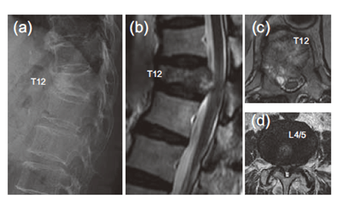
Fig. 1 Representative radiological images.
(a) A compression fracture with consolidation was found in the anterior part of the T12 vertebral body.(b) Sagittal magnetic resonance imaging(MRI) showing a diffuse low intensity area of the T12 vertebral body, indicating metastasis about to contact the posterior spinal cord. (c) Axial MRI showing a metastatic lesion predominantly in the right portion of the T12 vertebral body, that was about to contact the spinal cord predominantly in the right side.(d) Asymptomatic but severe stenosis was observed at the L4/5 level.
The primary operation was performed according to previous literature,[2]which involved en bloc resection of the T12 vertebral body followed by posterior fixation in the prone position with an operative time of 5:56 (h:min) and 3,800 mL of blood loss. An expandable cage(Stryker, Kalamazoo, MI) was installed during anterior column reconstruction followed by 2-above- 2-below pedicle screw(PS) fixation of the T10-11 to L1-2 levels with placement of cranial lateral hooks at the T9 level(Figure 2(a-d)). The postoperative X-ray showed a possible endplate violation at the caudal vertebrae. The posterior fixation range was determined considering a possible future adjacent segment disorder at the L4-5 level, which is generally considered when performing long floating fusion ending at the L4 or L5 level.[3]The patient started rehabilitation two days after the surgery with a brace on her trunk, and she had a good postoperative course without pain and paralysis.
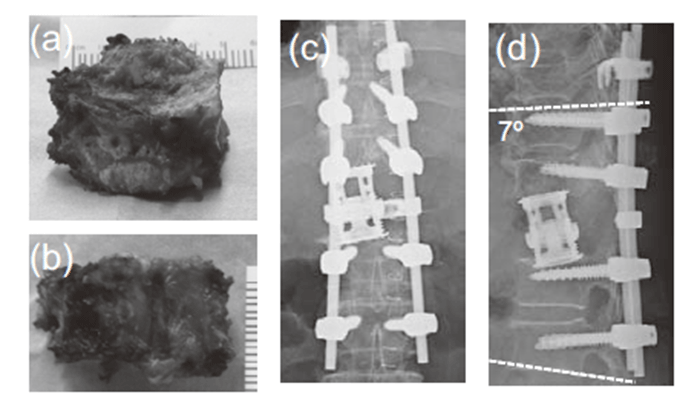
Fig. 2 Total en bloc spondylectomy of the T12 vertebral body.
(a, b) The vertebral body was resected in an en bloc manner.(c, d) The anterior column was reconstructed using an expandable cage with posterior fixation of 2-above-2-below fixation using cephalad transverse hooks at the T10 level. The kyphotic angle was 7° between the T10 and L2 levels.
Three weeks after primary TES, a follow-up radiograph showed progressive junctional kyphosis at the site of TES though the patient showed no related symptoms such as pain, numbness, and paralysis. Radiological evaluation also showed an adjacent vertebral fracture of the expanded, predominantly in the caudal vertebral body with marked subsidence. The cage had direct contact with the adjacent L1 PS, and the caudal L2 PS showed forced loosening. The kyphotic angle of T10-L2 increased from 7° to 15° with sagittal instability during flexion(Figure 3(a-c)). Over time, follow-up radiographs showed progressive kyphosis, which led to further collapse of the caudal component. Therefore, on the basis of these findings, we decided to perform revision enforcement surgery. In the prone position, the loose L2 PS was removed, and then additional posterior fixation was performed using L3-4- 5-S1followed by S2 alar-iliac(S2AI) screw fixation to achieve secure fixation. A median hook was also placed at the L2 spinous process to reinforce the compressive force of the L2 vertebral body in the cranial direction, and then a lateral interbody fusion cage(Medtronic, Minneapolis, MN) was inserted at the L1-2 level using the oblique lateral interbody fusion approach to gain more direct support in the right decubitus position. The kyphotic angle improved from 8° to 15° after revision surgery (Figure 4 (a, b)). After revision surgery, the patient started rehabilitation two days after the surgery with her blace on to fully get recovered in daily activities with no neurological symptoms, and returned to activities of daily living with maintained. A bony bridge between the T11 and L1 vertebral bodies developed 1.5 year postoperatively and was confirmed on a reconstructed computed tomography scan.
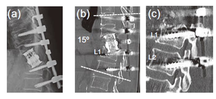
Fig. 3 Radiological images 3 weeks postoperatively.
(a, b) Fractures adjacent to the vertebral bodies caused by the expandable cage were predominantly found in the L1 vertebral body. Local kyphosis from T10-T2 was exacerbated from 7° to 15°, causing spinal kyphosis.(c) The void lesion above the L2 PS, which was forced by the compressive moment caused by the cephalad components, is noteworthy.
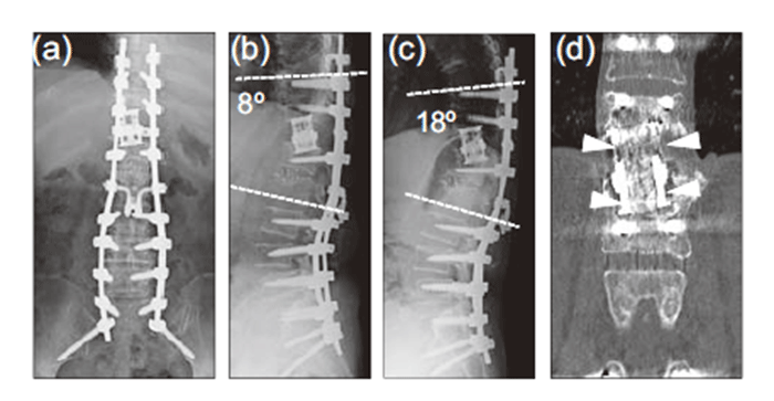
Fig. 4 Revision surgery performed 1 month after the primary surgery.
(a, b) Radiographs at 1.5 years postoperatively. After removing the L2 pedicle screw because of major loosening, additional posterior fixation was performed using L3-4-5-S1 PS and S2 alar-iliac screws. The median hook was added to reinforce the compressive force of the L2 vertebral body in the cephalad direction, and then the lateral interbody fusion cage was inserted at the L1–2 level using the oblique lateral interbody fusion approach to gain more direct support. The local kyphotic angle at T10-L2 improved to 8°(c), finally 18º with correction angle loss of 10° at the final observation(18 months after the surgery, d).(e) Post-operative CT scan showed massive bony fusion connecting the adjacent vertebrae(arrow heads).
The main reasons for primary implant failure in our case were as follows:(1) inappropriate installment of the expandable cage and(2) fixation with a relatively short range, i.e., 2-above-2-below fixation.
First, we should have considered that the patient was osteoporotic. Implant failure in osteoporotic patients has been reported in several previous reports, and one study explained the importance of endplate preservation in such patients.[4]In ordinary osteoporotic patients preoperatively, medications to improve bone density should be considered to acquire earlier bony fusion and less implant failure, and increase bone mineral density.[5,6]
TES is one of the effective surgical techniques for solitary spinal metastasis,[2]but it can also cause major instability after three-column resection.[7]Thus, there may be a good indication for using expandable cages to achieve efficient and immediate correction of spinal malalignment[7,8], which explains why we used an expandable cage in our case. However, expandable cage has some reported complications such as adjacentlevel vertebral body fractures after expandable cage reconstruction.[9]The current case had an adjacent vertebral body fracture, predominantly in the caudal endplate with marked subsidence. When using expandable cages, subsidence is almost unavoidable with a mean value of 1.4 mm,[10]which is sometimes required to maintain stable contact with the adjacent endplates.[7]However, in cases with weakened endplates, i.e., with excess intra-operative violation of endplates or osteoporotic patients, subsidence can cause too much intra-vertebral subduction. Early cage subsidence proposed in such cases causes a loss of tension within the stabilized construct, resulting in a decrease of the primary stability and an overload of the remaining load-bearing structures as seen in the current case.
Another risk of expandable cages is overdistraction [11]. There is no clear sign to warn the surgeon to stop expansion during surgery, as shown in the current patient with an osteoporotic vertebral body who underwent three-column resection. The combination of an anterior expansion force and posterior compression force should have worked as excess destruction forces to the adjacent vertebral bodies. Thus, in the current case, we should have expanded the cage to a less extent so that it would make contact with the bony endplate without excess distraction force since compressive posterior fixation would be performed later. Spinal shortening achieved by TES has important advantages such as increased spinal stability and increment in spinal cord blood flow, which were not observed in the current case. Overdistraction and marked subsidence of the cage should be avoided to achieve these advantages.
We should have also taken care of the range of posterior fixation. A previous biomechanical human cadaveric study indicated that posterior PS with 2-above and 2-below constructs can provide sufficient rigid fixation during thoracolumbar corpectomy.[12]The current patient underwent posterior L2 fusion on the basis of this evidence. As a result, this strategy was inadequate for anchoring the fused component; thus, we performed additional fusion of the iliac using the S2AI trajectory to ensure rigid fusion. We should have considered the patient's osteoporotic state in planning the primary posterior fusion range. Too short posterior fixation cannot counteract the imbalance due to muscle and ligament sacrifice, which in our case resulted in implant failure.
Surgeons also have to pay more attention if patients have a history of irradiation, which can cause other complications such as delayed incision healing and cerebrospinal fluid leakage[13], which is one of the reasons to perform surgery before radiation.
In conclusion, we experienced an implant failure with early subsidence of an anterior expandable cage with possible intra-opeartive violation on endplates and short posterior fixation in an osteoporotic patient with a solitary spinal metastasis. We performed revision surgery by adding posterior fixation to S2AI followed by using an anterior lateral intervertebral cage. Less intra-operative violation on endplates that can be achieved by adequate expansion, and sufficient long-segmental posterior fusion should have been considered in the current case.
Written informed consent was obtained from the patient for publication of this case report and any accompanying images. A copy of the written consent is available for review by the Editor-in-Chief of this journal.
The authors report no conflict of interest concerning the materials and methods used in this study or the findings specified in this paper.
The original datasets supporting the conclusions of this article are not available, because these are originally personal data in our facility.
TI, SuO, HK, YS, GO, TS, KI, KF, JS, YS, HK, KA SeO analyzed and interpreted the patient data. TI, SuO and SeO performed the surgeries. YK, JN, YM, YA, YE, TF, MK, MS and KT were major contributors in writing the manuscript. All authors read and approved the final manuscript.
We thank Dr. Akihiko Okawa at Chiba medical center for medical advices on the case, including preand post-operative treatment and surgical indication. Dr. Okawa, as well as all of the authors, has no fund to declare in the present case.
Address correspondence to Dr. Sumihisa Orita.
Department of Orthopaedic Surgery, Graduate School of Medicine, Chiba University, 1-8-1, Inohana, Chuou-ku, Chiba 260-8670, Japan.
Phone: +81-43-226-2117. Fax: +81-43-226-2116.
E-mail:sorita@chiba-u.jp
Abbreviations:TES: Total en bloc spondylectomy, T: Thoracic spine, L: Lumbar spine, PS: Pedicle screw, S: Sacrum, S2AI: S2 alar iliac
