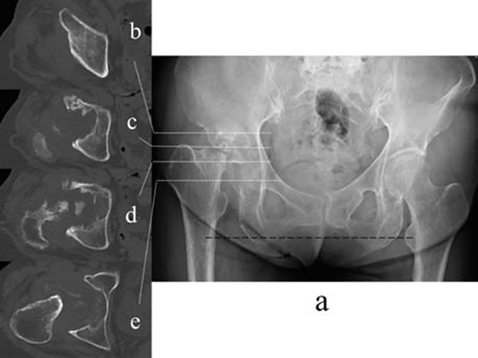
Fig. 1 Imaging features of the pelvis at the first visit.
Anteroposterior X-ray image shows severe joint destruction of the right hip with disappearance of the femoral head and the right femoral shortening followed by leg length discrepancy (horizontal reference line passing the left lesser trochanter in black broken line)(a). Computed tomography reveals destruction of the acetabulum at the: roof(b), joint line(b), center of the acetabulum(c), and tear drop(d).