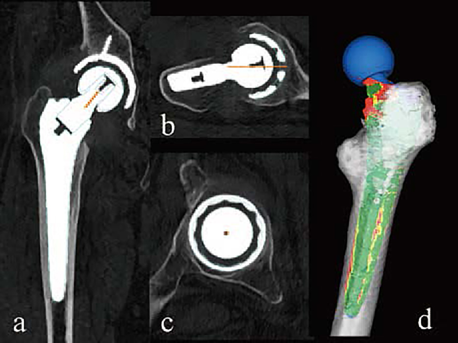
Fig. 3 Postoperative evaluation of implant alignment with three dimensional, CT-based software.
Coronal reconstruction(a), oblique axial reconstruction of the femoral neck(b), oblique sagittal reconstruction of the cup(c) shows 33° of cup radiographic inclination, 13° of radiographic anteversion, 15° of stem anteversion, and 27° of combined anteversion(postoperative templating in blue). Three dimensional, canal filling analysis indicates that the edges along the square stem fit well in yellow and red.