


Chiba Medical J. 95E:63-69,2019
doi:10.20776/S03035476-95E-4-P63
[ Chiba Medical Society Award Review ]
Hiroyuki Takaoka
Department of Cardiovascular Medicine, Chiba University Graduate School of Medicine, Chiba 260-8670.
(Received March 18, 2019, Accepted May 24, 2019, Published August 10, 2019.)
Since the introduction of electrocardiography-gating scan technology and multislice detectors in the field of computed tomography(CT), significant improvements in the noninvasive diagnosis of cardiovascular diseases, including coronary artery disease, have been achieved using CT in daily clinical practice. After the introduction of new-generation CT, we have been using cardiac CT not only in our daily clinical practice but also in several clinical studies. First, we focused on evaluating plaques of coronary arteries and reported the usefulness of CT in assessing plaque characteristics in patients with acute coronary syndrome. Next, we demonstrated the high diagnostic accuracy of significant coronary artery stenosis using new generation 320-slice CT even in patients with arrhythmia or tachycardia who were not appropriate candidates for cardiac CT before the introduction of the 320-slice CT. We also evaluated myocardial damage as late enhancement in left ventricular myocardium on CT and identified it as a marker of future cardiac events in patients with hypertrophic cardiomyopathy. We reported higher diagnostic accuracy of myocardial damage, on the new-generation 320-slice CT with new iterative reconstruction techniques in patients with non-ischemic cardiomyopathy. Further, we helped medical and postgraduate students with their cardiac imaging-related research. We also helped them win several prizes at medical conferences and write papers for worldwide medical journals in English. Here, we would like to introduce our contribution to the development of noninvasive diagnosis of cardiovascular diseases and to the cardiac imaging-related educational achievement for medical and postgraduate students.
Cardiovascular disease, Computed tomography, Coronary artery, Myocardial fibrosis, Cardiac imaging technology
Significant improvements have been noted in the noninvasive diagnosis of cardiovascular diseases in daily clinical practice since the introduction of electrocardiography(ECG)-gating scan technology and multislice detectors in the field of computed tomography (CT)[1]. Several studies have identified the high negative predictive value of CT in the evaluation of coronary artery stenosis; hence, CT has been widely used for this purpose[2]. Since the introduction of newgeneration CT at our institution, we have been using cardiac CT not only in our daily clinical practice but also in several clinical studies. We initially focused on evaluating coronary arteries using CT and then evaluated the utility of CT for assessing myocardial damage.
We also helped medical and postgraduate students in conducting cardiac imaging-related research, winning several prizes at medical conferences, and writing papers for medical journals. Through this paper, we would like to inform the readers about our contribution to the improvements in the noninvasive diagnosis of cardiovascular diseases on CT and to the cardiac imaging-related educational achievement for medical and postgraduate students.
In almost 70% patients with acute myocardial infarction, the responsible lesions on coronary arteries showed <50% stenosis upon invasive coronary angiography conducted before disease onset[3]. Recently, sudden plaque rupture and subsequent coronary artery occlusion by thrombus was recognized as the major mechanism underlying acute coronary syndrome(ACS) onset[4]. Characteristics of the plaques of the coronary arteries of patients with ACS were identified previously using IVUS; fatty components and positive remodeling or spotty calcification were noted as the typical characteristics of unstable high-risk plaques causing ACS[5], and those characteristics were also detectable with CT(Fig. 1A, 1B and 1C). Evaluation of the plaque characteristics was expected to be useful for predicting future coronary artery events. Therefore, we aimed to investigate the utility of CT for evaluating the characteristics of plaques of coronary arteries in patients with ACS.
A total of 31 consecutive patients who were diagnosed with ACS and underwent both cardiac CT and invasive coronary angiography(ICAG) with IVUS were retrospectively selected[6]. The CT and IVUS images of the culprit lesions causing ACS(culprit lesions) were compared. CT values of the culprit lesions diagnosed as soft plaques using IVUS(n=6; 32.9± 8.7 Hounsfield Unit〈 HU〉) did not significantly differ from those diagnosed as thrombi(n=18; 43.2±10.7 HU; p=0.268); further, the values were significantly lower than of those diagnosed as fibrotic plaques, which were also diagnosed using IVUS(n=7; 82.5± 22.6 HU; both p <0.01). There were no significant difference of the percentages of the patients with calcifications(67.7%), spotty calcifications(61.3%), and positive arterial remodeling(58.1%) between in IVUS and CT images(67.7% vs 58.1%, 61.3% vs 51.6%, and 58.1 vs 74.2%, respectively; p=0.43, 0.44 and 0.18). Therefore, we concluded that CT was useful for detecting the characteristics of the culprit lesions in patients with ACS. We won the Young Investigators Award at the 49th Japanese College of Angiology in October 2008.
As I mentioned, evaluation of plaque components, not stenosis, of coronary arteries was expected to be useful for prediction of future ACS events[3-7]. Therefore, we aimed to investigate the utility of evaluating the characteristics of the plaques of coronary arteries on CT for predicting future cardiac events.
We selected 195 patients who underwent coronary CT performed using 8 or 16-slice CT between November 2002 and December 2003 at our institution [8]. The included patients did not have significant coronary artery stenosis on CT and history of myocardial infarction or coronary bypass surgery. We evaluated the patients for the presence of non-calcified plaque(NCP) or mixed plaque(MP) on CT. All patients had a normal sinus rhythm during CT. The relationship between their ACS events after cardiac CT and the presence of NCP or MP on CT were evaluated. All the patients were retrospectively followed up for 82±40 months after CT. Only two patients demonstrated ACS during the followupperiod. Receiver operating characteristic(ROC) analysis of the numbers of NCPs or MPs of coronary arteries detected on CT was performed for evaluating ACS onset. Based on ROC analysis, number of NCP or MP > 0 was the best cutoff value; the sensitivity and specificity were 67% and 80%, respectively. The area under the ROC curve was 0.604. Patients with > 0 NCP or MP on CT(n=48) were older and showed a high prevalence of diabetes mellitus, dyslipidemia, history of smoking, and oral intake of a small amount of aspirin. There was no significant difference in ACS occurrence between the patients with > 0 NCP or MP and the other patients. However, the higher population of intake of aspirin in patients with > 0 NCP or MP, the small number of the patients who had ACS and the difference of prevalence ratio of coronary risk factors might have significantly influenced this result. In this retrospective analysis, we would like to set a longer follow-up period for all patients. Therefore we selected patients who underwent cardiac CT using an old CT scanner, and the plaque component classification was limited to NCP or MP. This was the major limitation of this research.
We presented our findings and subsequently received the Japanese College of Angiology Award at the 54th annual meeting of the Japanese College of Angiology in 2013. We also received the Japan Heart Foundation Research Grant(2 million yen).
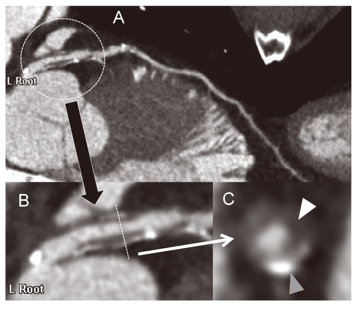
Fig. 1
Typical plaque image with positive remodeling, spotty calcification, and low attenuated lesion of a coronary artery in a curved planner reconstructed image on CT(A). The plaque image is magnified (B), and spotty calcification(gray arrow heads) and low attenuated plaque(white arrow heads) were obvious in a short axial image(C).
Before the introduction of the 320-slice CT (Aquilion One/ Cannon Medical Systems, Otawara, Japan; Fig. 2A), patients with unstable HRs including arrhythmia were not eligible candidates for coronary CT because of the decrease in the image quality of the coronary arteries on CT(Fig. 2B). The 320-slice CT was expected to overcome this limitation of cardiac CT; the 320-slice CT has a 16-cm craniocaudal coverage and can produce high quality images, without stepping artifacts, of the entire coronary arteries in a single heartbeat(Fig. 2C). Therefore, we evaluated the utility of 320-slice CT for evaluating coronary artery stenosis in 106 consecutive patients, including those with arrhythmias or higher HRs(n=44) who were suspected of having coronary artery disease and underwent cardiac CT and invasive coronary angiography within 3 months [9]. Segment-based-analysis revealed a sensitivity and specificity of detection of significant(>75%) coronary artery stenosis of 86% and 98% in patients with normal sinus rhythm and lower HRs[<65 beats per minute(bpm)] and 68% and 99% in patients with higher HRs(>64 bpm) or arrhythmia(p=0.101, or 0.174), respectively. The diagnostic accuracy of significant coronary artery stenosis was preserved even in patients with arrhythmias or higher HRs who were not appropriate candidates for cardiac CT before the introduction of 320-slice CT.
In the beginning, the radiation dose for cardiac CT was higher than that for conventional plain chest CT, therefore it had been concerned by many physicians. [10]. Previously, retrospective ECG-gating scanning (radiation is exposed during all cardiac phases) was necessary for cardiac CT acquisition; however, recently, a prospective ECG-gating(radiation is exposed during limited cardiac phases, mostly only during the middiastolic phases because of the minimum motion artifact) technique was established for reducing the total radiation dose for cardiac CT.
We would like to decrease the total cardiac CT scanning time for reduction of radiation exposure without decreasing the diagnostic accuracy of significant coronary artery stenosis. We investigated the best scanning time of cardiac CT for the precise evaluation of significant coronary artery stenosis on CT. We retrospectively analyzed 77 consecutive patients who underwent both cardiac CT with retrospective ECGgating and invasive coronary angiography within 3 months and classified them into 2 groups based on HR during CT scanning(group 1, 44 patients with HR< 66 bpm; group 2, 35 patients with HR>65 bpm)[11]. CT images were reconstructed at every 5% increased of whole RR-interval. Three methods were used to detect significant coronary artery stenosis >50%: 1) using only 75% of the data named virtual prospective ECGgating without padding, 2) using 70%-100% of the data of group 1 or 35%-100% of the data of group 2 named as virtual prospective ECG-gating with padding, and 3) using all-phase data named retrospective ECGgating. There were no unevaluable segments in all three methods in group 1. However, the percentage of unevaluable segments was significantly higher with the virtual prospective ECG-gating without padding (13.6%±27.9%) than with the virtual prospective ECG-gating with padding (0.7%±3.1%) and the retrospective ECG-gating (0.7%±3.1%) (both p=0.012) in group 2. In each group, the diagnostic accuracy of significant coronary artery stenosis on CT did not differ among the three methods, using only evaluable segments. Virtual prospective ECG gating at 75% of the R wave-to-R wave(RR)-interval without padding had significantly more unevaluable segments than the virtual prospective ECG-gating with padding and retrospective ECG-gating only in subjects with HR>65 bpm. We concluded that the total cardiac CT radiation dose could be reduced in patients with lower HR and that diagnostic accuracy was preserved even with the short scan time.
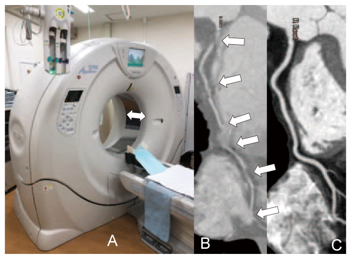
Fig. 2
320-slice Computed Tomography(CT) has a 16- cm craniocaudal coverage, which is indicated by a white arrow(A), and it allows us to obtain clear images of coronary arteries without any stepping artifacts even in patients with frequent arrhythmia. Two typical different curved planner reconstructed images of the right coronary artery on CT in the same patient with atrial fibrillation on 16-slice CT (B) and 320-slice CT(C). Stepping artifact was obvious on 16-slice CT(white arrows) because of its short craniocaudal coverage; however, the artifacts were diminished on 320-slice CT.
Cardiac magnetic resonance imaging(MRI) is a useful modality for detecting myocardial fibrosis(MF) as LE(Fig. 3A)[12]. Evaluation of LE is useful in the prediction of future cardiac events and the differential diagnosis of several myocardial diseases[13]. Because cardiac MRI requires a highly skilled radiologic technologist and a longer acquisition time, the number of cardiac MRIs performed matched only 10% of the number of cardiac CTs performed in our country[14]. Further, CT has reportedly been used for detecting myocardial damage; however, the CT image quality of LE was not suitable for daily clinical use[15]. CT has recently been mechanically improved, and we would like to clarify the utility of cardiac CT for detecting myocardial damage as LE.
We retrospectively analyzed 56 consecutive patients who were suspected of having myocardial disease or cardiac tumor and underwent cardiac 16-slice CT and 1.5-Tesla MRI within 2 months [16]. We also evaluated the diagnostic accuracy of cardiac CT for detecting myocardial damage as LE in LV myocardium(LVM) In 31 patients, LE was detected on both CT and MRI; it was also detected in 192 and 197 LV segments on CT and MRI, respectively. The sensitivity, specificity, and diagnostic accuracy of LE on CT in comparison with MRI as the gold standard were 90%, 89%, and 89% in the patient-based analysis and 67%, 92%, and 87% in the segment-based analysis(using the 17-segments model, AHA), respectively. Here, we clarified the utility of CT for detecting LV myocardial damage.
We compared the influence of non-sustained ventricular tachycardia(NSVT) and LE in LVM on CT, mean MF, and risk stratification for major adverse cardiac events(MACE) in patients with hypertrophic cardiomyopathy(HCM) without obstructed coronary arteries. We selected 45 consecutive patients with symptomatic HCM who underwent cardiac CT, transthoracic echocardiography and 24 hours ECG monitoring within 12 months and had no obstructed coronary arteries(>50%)[17]. NSVT was detected in 23 patients, and no significant differences of MACE occurrence were observed between HCM patients with and without NSVT at each time point and when the entire follow-up period was compared using the Kaplan- Meier and log-rank tests (p=0.188). Significant differences of MACE occurrence between HCM patients with and without MF were observed at each time point and when the entire follow-up period was compared by Kaplan-Meier analysis and log-rank test(p=0.049). We revealed the utility of detecting LE in LVM on CT for predicting future cardiac events in patients with HCM.
Recently, FIRST, a new iterative reconstruction technique, was equipped with the latest 320-slice CT for use in daily clinical practice. FIRST is a useful technique for obtaining high-quality CT images under low radiation and contrast acquisition[18]. Therefore, we aimed to evaluate the utility of a new generation 320 slice CT with FIRST for detecting myocardial damage as abnormal LE in LVM on CT(Fig. 3B). We retrospectively analyzed 88 patients without ischemic heart disease who underwent cardiac CT using various scanners and 1.5 or 3 Tesla MRI within 3 months [19]. We also evaluated the diagnostic accuracy of a new generation 320-slice CT with FIRST for detecting myocardial damage as LE in LVM in comparison with the old CT scanners with filtered back projection (FBP), an old reconstruction method. We classified all 88 patients into 3 groups based on the CT scanner used for cardiac CT(group 1, 52 consecutive patients who underwent 16-slice CT with FBP at 140 kV tube voltage and an average tube current of 337±20 mA; group 2, 18 consecutive patients who underwent 320-slice CT with FBP at 120 kV tube voltage and an average tube current of 255±106 mA; group 3, 18 consecutive patients who underwent new-generation 320-slice CT with FIRST at 80 kV tube voltage and a tube current of 800 mA). On patient-based analysis, no significant differences were observed between the 3 groups. On segment-based analysis, Sensitivity, specificity, PPV, NPV, and overall accuracy of detection of LE on CT compared to MRI were 65, 92, 68, 91, and 87%, respectively in Group 1, 67, 94, 66, 95, and 91% respectively in Group 2, and 73, 97, 85, 95, and 93%, respectively in Group 3. Specificity and overall accuracy were significantly higher in Group 3 than in Group 1(both P<0.01). PPV was significantly higher in Group 3 than in both Group 1 and 2(both P<0.05). We clarified the utility of combination of new generation CT using lower kV, higher mA and FIRST for detection of myocardial damage on CT.
Takaoka H, received the young investigators’ award(YIA) at the 27th annual meeting of the Japanese Society of Cardiovascular Imaging(Jan 2017), the clinical research award at the 242nd Kantokoshinetsu regional meeting of the Japanese Circulation Society (JCS)(June 2017), and a research grant from the Tsuchiya Memorial Foundation for my research on the evaluation of LE in LVM on CT using FIRST.

Fig. 3
Typical late enhancement images on magnetic resonance imaging (MRI) (A) and CT (B) in a patient with paroxysmal atrial fibrillation and myocardial damage in the left ventricular myocardium caused by an unknown etiology.
We helped some medical students receive a total of five awards at several medical conferences(the annual meetings of the Japanese Society of Internal Medicine and Kantokoshinetsu regional conferences of JCS), four awards from the President of Chiba University, and 3 Encouragement Awards of Scholarship Program of School of Medicine, Chiba University. We also helped medical students write seven papers for worldwide medical journals in English[20-26]]. Our postgraduate students received two YIAs at the Kantokoshinetsu regional conferences of JCS and one YIA at the annua meeting of the Myocardial Biopsy Society. Based on our abovementioned experience, we would like medical students to continue motivating for conducting clinical research and help them to be brilliant medical researchers who will work at the global medical stage.
We contributed to the development of noninvasive diagnosis and clinical research of cardiovascular diseases using cardiac CT and to the cardiac imagingrelated education of medical and postgraduate students.
I would like to express my sincere thanks to my mentors, colleagues and collaborators, especially to Prof. Yoshio Kobayashi, Prof. Issei Komuro, Dr. Iwao Ishibashi, Dr. Nobusada Funabashi, Dr. Masae Uehara and Dr. Koya Ozawa. I also want to express my thanks to Prof. Koichi Sano for critically reviewing this article. This work was partially supported by the TSUCHIYA MEMORIAL MEDICAL FOUNDATION (Grant no. J17KF00167).
HT was responsible for writing the manuscript and preparing the figures. Written informed consent to publication was obtained from the patients, whose cardiac CT and MRI images were used in all figures.
The author declare that they have no conflicts of interest, either financial or non-financial, with the contents of this article.
Address correspondence to Dr. Hiroyuki Takaoka.
Department of Cardiovascular Medicine, Chiba University Graduate School of Medicine, 1-8-1, Inohana, Chuou-ku, Chiba 260-8670, Japan.
Phone: +81-43-226-2555. Fax: +81-43-226-2557.
E-mail:tapy21century@yahoo.co.jp
