


Chiba Medical J. 95E:79-84, 2019
doi:10.20776/S03035476-95E-6-P79
[ Case Report ]
Hiroshi Yoshimura1,2), Tomoo Kise1), Shigeru Fukuyama1),
Masatsugu Uehara1), Keiji Akamine1,3), and Naoki Shimizu2,4)
1 ) Department of Pediatric Nephrology, Prefectural Okinawa Nanbu & Children’s Medical Center, Haebaru Town, Okinawa 901-1193 .
2 ) Department of Pediatrics, St. Marianna University School of Medicine, Kawasaki City, Kanagawa 216-8511 .
3 ) Department of Pediatric Nephrology, Tokyo Metropolitan Children’s Medical Center, Tokyo 183-8561 .
4 ) Department of Pediatric Emergency & Critical Care Medicine, Tokyo Metropolitan Children’s Medical Center, Tokyo 183-8561 .
(Received August 7, 2019, Accepted September 5, 2019, Published December 10, 2019.)
Kidney transplantation (KTx) in infants (body weight [BW] <10 kg and/or body surface area [BSA] <0.5m2) is an accepted treatment option for chronic kidney disease in children and has demonstrated excellent short-term graft survival. However, follow-up studies longer than five years are still lacking. We experienced a case of an infant (BW 9.2 kg, BSA 0.44m2) with an adult-sized kidney (ASK) transplant (donor-recipient height ratio: 2.24) which resulted in graft loss at 9.7 years after the transplant secondary to biopsy-proven chronic allograft ischemia (CAI). Intensive maintenance fluid supplementation failed to prevent ongoing and long-standing graft hypoperfusion. While the decrease in the estimated glomerular filtration rate (eGFR) was irregular, the progression of chronic changes seen in the graft biopsies was linear, both in early- and late-phase KTx. CAI in significantly sizemismatched ASKs transplanted into infants can promote late graft failure within ten years. Since serial evaluations of renal histology were shown to be a better indicator of renal graft outcomes throughout the post-transplant course, transplant surveillance biopsies beyond the early phase of KTx may assist in elucidating the natural course of CAI in ASKs transplanted into infant recipients.
Kidney transplantation, infant, adult-sized kidney, chronic allograft ischemia, long-term graft survival
Kidney transplantation (KTx) in infants (body weight[BW]<10 kg, and/or body surface area[BSA]<0.5m2) starting dialysis in the neonatal period is an emergency intervention for unstable efficacy of dialysis and possible loss of peritoneal/vascular access for dialysis[1,2]. Even among this patient population, recent, sophisticated, pre- and post-transplant, medical, surgical, and multi-disciplinary interventions have impoved short-term renal allograft survival within five years after KTx[3-5]. In contrast, several studies reported an early phase of chronic allograft ischemia (CAI) in adult-sized kidneys (ASKs) transplanted into small children (BW <15 kg, and/or BSA <0.75m2) due to the considerable donor-recipient size mismatch, represented by donor-recipient BSA or height ratio ≥1.8 to 2.0[6-10]. Among these studies, two demonstrated a correlation between the increasing degree of sizemismatch and increasing evidence of ischemic changes in allograft biopsies developing as early as one year post-transplant and progressing steadily over the next two years[8,11]. It is theoretically predicted that more rapid progression of ischemic change and shorter graft survival will be exhibited in more size-discrepant KTx, i.e., ASK transplantation in‘ infants’, the further special group of small children, with donor-recipient BSA or height ratio ≥2.2 to 2.5; however, the actual longitudinal effect of CAI on graft survival in this special KTx category has yet to be described[12]. We herein report CAI causing late graft loss relatively sooner(<10 years) in an infant with an ASK transplant over a decade of continuous observation. A series of scheduled and clinically indicated graft biopsies over the time period enabled more accurate monitoring of graft function than the serum creatinine(SCr)-based estimated glomerular filtration rate (eGFR).
A female infant was born at 35 weeks of gestation with a birth weight of 2846 g via urgent cesarean section for progressive oligohydramnios, which presented immediately prior to delivery. Her clinical course was complicated by severe asphyxia due to dry lung syndrome, total anuria, severe hypertension, and abdominal compartment with bilateral enlarged kidneys. The patient required ventilator support, peritoneal dialysis (PD), and bilateral nephrectomy. The histology of her native kidneys and an examination of her parents’ bilateral kidneys confirmed the diagnosis of autosomal recessive polycystic kidney disease.Intensive, continuous cycling PD and aggressive nutritional support enabled growth within the minimally acceptable range. At around age 12 months, she experienced peritoneal membrane failure, multiple thromboses in the central veins, and subsequently, underdialyzed status, which prompted us to perform a living related KTx from her mother. The patient underwent transplant surgery at age 14 months. Her BW at the time was 9.3 kg (+0.3 standard deviation [SD]), her BSA was 0.44m2, and her height was 68.4 cm (-2.1 SD). Her mother’s height was 153.6 cm, giving the donor-recipient height ratio of 2.24. The operation was successful with immediate graft function, and the patient was placed on quadruple immunosuppression consisting of basiliximab, tacrolimus, mycophenolate mofetil, and methylprednisolone. To minimize hypovolemia, a major predisposing factor of renal allograft ischemia in this BW/BSA size group, a total daily fluid intake of 2500-3000 mL/m2/day with a sodium intake of at least 10 mEq/kg/day via nasogastric tube was maintained. The volume of fluids were strictly titrated according to her clinical hydration status and Scr levels as previously recommended[8,13]. To avoid dehydration, adequate intravenous isotonic fluids were supplemented immediately at every occurrence of common airway or gastrointestinal viral illness. This fluid management was continued for more than six years until stage G4 chronic kidney disease (CKD), i.e., the eGFR of 15- 20 mL/min/1.73m2, developed, at which time dialysis was begun again. Throughout the course, she experience no dehydration-associated hypotension or medicationor graft dysfunction-related hypertension until the development of Stage G4 CKD. Her SCr levels were stable at 0.29-0.31 mg/dL until three years post-KTx. Her eGFR, calculated using a formula developed for Japanese children based on SCr and height[14], was also stable. However, at 9.7 years post-KTx her eGFR fell to zero(Fig.1). She underwent a graft biopsy at one hour, one year, two years, and five years post- KTx as surveillance, and at three and six years post- KTx due to an asymptomatic increase in the SCr. Surprisingly, all the histological findings of the biopsies during the entire course were nearly identical, showing predominantly interstitial fibrosis and tubular atrophy (IFTA) disproportionate to glomerular structural changes and indicative of ischemic nephropathy[15] (Fig. 2). While the eGFR remained at nearly the same level for a certain period and then dropped abruptly in a stepwise manner, the histologically preserved area of the core biopsy specimen (the actual functioning area) declined linearly from the early to late post-KTx period, i.e., IFTA progressed steadily from ci 0 to ci 3 and ct 0 to ct 3 of the Banff Classification[16], showing a considerable difference between the graft histology and the eGFR. The degree of IFTA change was far greater than the GFR decrease not only between years 2 and 3, but also between years 5 and 6 post-KT(Fig.1). Serial histological findings and blood/urine tests denied the following possible causes of chronic renal allograft injury other than CAI: T cell-/antibody-mediated rejection, acute/chronic calcineurin inhibitor toxicity, de novo renal diseases, and latent viral infections caused by cytomegalovirus/Epstein-Barr virus/polyomavirus. At each biopsy, the ongoing ischemic process prompted us aggressively to reinforce the fluid regimen. After all our efforts failed to slow the progression of this process, dialysis was resumed at 7.5 years post-KTx, and the patient became totally anuric two years after resuming dialysis.
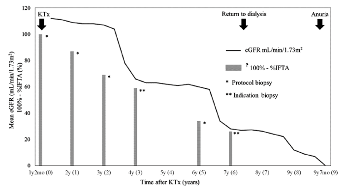
Fig.1 Course of eGFR changes and the histologically preserved(morphologically functioning) area in the core biopsy specimen(100%-%IFTA)
Numbers within the parenthesis next to the labels on the horizontal axis indicate post-transplant years.
KTx: kidney transplantation, eGFR: estimated glomerular filtration rate, IFTA: interstitial fibrosis and tubular atrophy, : %IFTA indicates the proportion of the graft core biopsy specimen showing interstitial fibrosis and tubular atrophy.
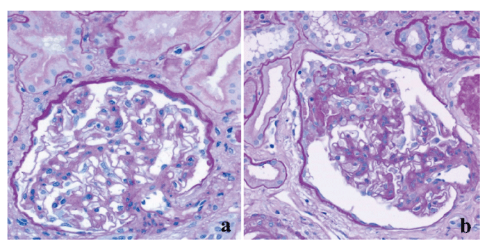
Fig.2 Two graft biopsies at one year(a) and three years (b) post-transplant (Periodic acid-Schiff[PAS] stain, magnification ×400)
Both show chronic ischemic nephropathy characterized by interstitial fibrosis and tubular atrophy (IFTA) with relatively preserved glomerular structures. IFTA, already presenting at one year post-transplant, progressed significantly at three years post-transplant, while eGFRwas unaffected during the period between the two time points.
This patient’s course provided two important clinical suggestions. First, CAI in significantly size-mismatched ASKs transplanted into infants can produce progressive loss of graft function within ten years post-KTx. Second, a renal histology is a better tool for assessing and predicting long-term renal allograft function than the eGFR throughout the post-KTx course.
First, continuous hypoperfusion of ASK allografts in infants (<10 kg, BSA <0.5m2) can be not only an important cause of chronic renal allograft injury, but also give rise to earlier late graft loss within ten years after KTx. As a mechanism of ischemic allograft dysfunction, previous studies demonstrated that ASKs transplanted into small children (<15 kg, BSA <0.75m2) resulted in inadequate blood flow, leading to the development of adaptive hypotrophy and failure to increase in size parallel to the patient’s maturation; histological results demonstrated IFTA at three months post-KTx which gradually progressed over one to two years[8,10,11,13,17-19]; however, given the shortterm (<5 years post-KTx) nature of the results and no differentiation between infants and small children in these studies, the impact of CAI on long-term (>5 years post-KTx) ASK allograft function in infants has yet to be clarified. The most recent studies with a large cohort have demonstrated improved ten-year graft survival rates of 74.0-81.1% in KTx in small children, which were equivalent to the results in older pediatric recipients[3,5,6,20]. Nonetheless, these reports did not specify the detailed recipient sizes or etiologies of graft failure accounting for approximately 20-25% of the graft loss cases; infants with CAI would likely have been a significant proportion of the graft loss population. Two studies demonstrated a mean rate of eGFR decrease in small children with KTx of 70-75 (ml/min/1.73m2), 55-60, and 40-45, at one, five, and ten years post-KTx, respectively. However, since the data on infants were not differentiated from those on small children[6,7], little is known with regard to the eGFR decrease rate in infants. One study reported that a donor-recipient height ratio of more than 2.2, mostly seen in ASKs transplanted into infants, was a high risk factor for graft loss within ten years[20]. This finding may corroborate the findings of our study that CAI may cause earlier late graft loss within a decade of transplantation in a case of infantile ASK KTx. In this case, the rate of eGFR decrease may be proportional to, or even exponentially greater, than the degree of the size mismatch.
Second, the renal histology of the interstitial fibrosis and tubular atrophy (IFTA) was a more sensitive and accurate indicator of actual renal function than the eGFR, both in the earlier and later post-KTx phases (>5 years). Two single center reports based on twoand five-year protocol graft biopsies in pediatric KTx demonstrated a fairly modest but statistically significant correlation between IFTA progression and eGFR decrease[9,21]; however, apart from the short-term nature of this finding, the authors were also concerned that the statistical power of their results was too weak to allow reliable clinical interpretation. Another shortterm study showed the superiority of surveillance graft biopsy findings to the eGFR by evaluating the actual functional status of pediatric KTx allografts[22]; later, the same authors suggested the need for long-term, serial surveillance biopsies for size-mismatched ASKs in infants and small children[23]. To date, more than 60% of pediatric KTx centers undertake only indication biopsies at the time of the abrupt eGFR decline during the follow up, much debating long-term benefits, timing, and safety of surveillance (protocol) biopsies; its’ timing and duration, if advocated, also vary depending upon the individual KTx centers, accordingly. However, several recent reports have demonstrated some evidence of improved outcomes via longitudinal biopsies with prescheduled points in time from several months to a dacade after KTx regardless of the recipients’ conditions, detecting modifiable conditions: subclinical acute cellular rejection (3 to 6 months after KTx), acute and chronic antibody-mediated rejection (a year to a decade after KTx), hypertension (a year to a decade after KTx), and chronic dehydration (as seen in this case report, 3 months to a decade after KTx) [24,25]. In our report, IFTA progression continued to be steady and predictable, while the eGFR decrease continued to be unstable and unpredictable for a decade. This finding emphasizes the importance of long-term histopathological evaluation via surveillance biopsy for ASK KTx in infants. Suggested timing of protocol biopsies in selected pediatric KTx recipients of the high risk group both for immunologic and non-immunologic graft loss may include time points of 1 hour (for baseline findings), 3 to 6 months, 1 year, 3 years, 5 years, 7 years and a dacade after KTx.
In conclusion, significantly size-mismatched ASK KTx in infants may lead to progressive chronic ischemic renal allograft injury as a late complication. Histopathological examination of IFTA may be superior to the eGFR as an indicator of allograft function immediately after a transplant and in succeeding years. Further studies are needed to determine whether distinctly size-mismatched ASK infant recipients manifest an earlier decline in graft function secondary to CAI and whether long-term protocol biopsies beyond five years post-KTx may help elucidate the natural course of CAI in ASK KTx in infants since a renal histology may an appropriate tool for evaluating graft function.
HY, TK, SF, MU, and KA managed the patient. HY, TK, SF, and MU contributed to writing this report. NS oversaw the writing process and contributed comments on the manuscript. Informed consent was obtained from the family for the preparation and publication of this report, both of which were also approved by the Institutional Review Board of Prefectural Okinawa Nanbu and Children’s Medical Center.
The authors are indebted to Iwao Nakazato of the Prefectural Okinawa Nanbu and Children’s Medical Center and Michio Nagata of the University of Tsukuba School of Medicine, for interpreting the graft renal biopsies. Authors also thank Mr. James Robert Valera for his assistance with editing the manuscript.
The authors declare that they have no conflicts of interest, either financial or non-financial, with regard to the content of this article.
Address correspondence to Dr. Hiroshi Yoshimura.
Department of Pediatrics, St. Marianna University School
of Medicine, 2-16-1, Sugao Miyamae-Ku, Kawasaki City,
Kanagawa 216-8511, Japan.
Phone: +81(-0)44-977-8111. Fax: +81(-0)44-976-8603.
E-mail:yoshimura.hiroshi@gmail.com
