


Chiba Medical J. 99E:27-37, 2023
doi:10.20776/S03035476-99E-3-P27
〔 Original Article 〕
Yasunori Matsumoto1), Masayuki Kano1), Hideki Hayashi1)
Hiroshi Suito1), Koichi Hayano1), Yoshihiro Kurata1), Ryota Otsuka1)
Hiraku Ono2), Koutaro Yokote2), and Hisahiro Matsubara1)
1) Department of Frontier Surgery, Chiba University Graduate School of Medicine, Chiba 260-8670.
2) Department of Endocrinology, Hematology and Gerontology, Chiba University Graduate School of Medicine, Chiba 260-8670.
(Received October 12, 2022, Accepted March 8, 2023, Published June 10, 2023.)
【Background】The association of liver dysfunction with morbid obesity has been speculated, which can be improved to some extent by metabolic surgery as represented by laparoscopic sleeve gastrectomy (LSG). However, the postoperative liver damage due to surgical procedures can sometimes be serious. Therefore, understanding the risk of perioperative liver damage and the positive effects of LSG on liver function is necessary.
【Materials and methods】A total of 46 morbid obesity cases that underwent LSG at Chiba University Hospital between 2010 and 2021 were evaluated for patient background, surgical factors, liver function and liver damage course and analyzed for perioperative liver damage risk factors.
【Results】The liver to spleen ratio (LS ratio) in plain computed tomography (CT) , an index that reflects liver fattening, significantly reduced 1 year after LSG compared to baseline values (P < 0.0001) , but without improvement in Fib-4 index, an indicator of liver fibrosis (P = 0.28). Liver damage (≧CTCAE grade2) was observed in 28.3% (13/46) of patients 3 days after surgery, with one patient requiring a platelet transfusion. The risk factors of perioperative liver damage include high Fib-4 index (P = 0.016) , high visceral fat subcutaneous fat ratio (VS ratio) (P = 0.049) , and long surgical duration (P = 0.023) using the multivariate logistic regression analysis.
【Conclusions】Avoiding perioperative liver damage to perform appropriate preoperative evaluation, including blood and imaging tests, and aiming to shorten the surgical duration is important for patients with morbid obesity undergoing LSG. LSG for morbid obesity shows hepatic fattening improvement with weight loss, but the effect on liver fibrosis should be examined including a longer course.
LSG, obesity, surgery, NASH, NAFLD
Obesity is a leading public health problem worldwide. In Japan, according to the statistics of the Ministry of Health, Labor, and Welfare in 2019, 33.0% of males over 20 years old and 22.3% of females are overweight with a body mass index (BMI) of 25 kg/m² or more, and the number of males is particularly rising. Obesity with a BMI of 30 kg/m2 or higher is found in 4.6% of adults, and the combined suspicion and reserve group for metabolic syndrome is estimated as 31.9% (The National Health and Nutrition Survey in Japan, 2019. https://www.mhlw.go.jp/content/001066903. pdf). The prevalence of non-alcoholic fatty liver disease (NAFLD) and non-alcoholic steatohepatitis (NASH) has considerably increased over the last few years. NAFLD is currently the most common cause of chronic liver disease in developing countries. More recently, experts have agreed that NAFLD does not reflect the current knowledge and have proposed metabolic (dysfunctional) -associated fatty liver disease (MAFLD) as a more appropriate umbrella term[1].
The spread of laparoscopic sleeve gastrectomy (LSG) for obesity also leads to an increased number of surgical cases for patients with obesity having liver dysfunction. The efficacy of LSG on NASH, NAFLD, or MAFLD with severe obesity was reported[2,3]. The improved effect of LSG on fatty liver, especially the liver to spleen ratio (LS ratio) in plain computed tomography (CT) was reported[4]. However, data on the effect of LSG on fibrosis or long-term liver function are insufficient and controversial. Perioperative liver damage in laparoscopic gastrectomy is often considered transient, but can sometimes be severe. The left lobe of the liver may be enlarged, and laceration, hematoma, or necrosis may develop in the liver due to retraction. Therefore, the types of retractors, etc. in LSG are being discussed to avoid these problems[5]
Here we discuss the risk of perioperative liver damage in LSG and the effect of LSG on mid- to longterm liver function.
This study was approved by the ethical committee of Chiba University and was conducted with 46 patients who underwent LSG due to obesity at Chiba University Hospital between September 2010 and August 2021. Patients’ clinical findings, surgical findings, and postoperative medium- to long-term course, as listed below, were retrospectively analyzed.
The short to long time changes in liver fattening, fibrosis, and damage after LSG are analyzed. Risk factors, such as clinical background and surgical factors, are assessed by comparing the groups with and without perioperative liver damage in LSG. Consent for this study in 46 cases was given on an opt-out basis. In addition, consent for the case report was obtained verbally.
Age, sex, height, weight, comorbidity (hypertension[HT], hyperlipidemia[HL], diabetes mellitus[DM], and sleep apnea syndrome SAS) , laboratory data (aspartate aminotransferase[AST], alanine aminotransferase[ALT], lactate dehydrogenase[LDH], gamma-glutamyltransferase[GGT], total bilirubin[T-Bil], direct bilirubin[D-Bil], albumin[Alb], platelet[Plt], total cholesterol[TC], HDLcholesterol[HDL-C], LDL-cholesterol[LDL-C], triglycerides[TG], C-reactive protein[CRP], and prothrombin time[PT]) , image findings (CT value of the liver and spleen, visceral fat area, and subcutaneous fat area) , operative time, blood loss, complication, hospital stay, and calculated scores, such as BMI, Child-Pugh score, NAFLD fibrosis score, Fib-4 index, LS ratio in plain CT, and visceral to subcutaneous fat area ratio (VS ratio).
The LS ratio in plain CT was used as an index of liver fattening. Fib-4 index calculated from age, AST, ALT, and Plt was used as an index of liver fibrosis. NAFLD fibrosis score, also an indicator of liver fibrosis, was calculated from age, BMI, AST, ALT, Plt, and Alb.
Perioperative liver damage is defined as liver enzymes of more than 3 times the upper limit of the reference value on postoperative days 3 and Grade 2 or more, based on Common Terminology Criteria for Adverse Events (CTCAE) ver.5.0.
A multidisciplinary conference was held to determine surgical indications. Medical inpatient treatment was performed for 2 weeks and surgical readmission was performed 2 days before surgery after temporary home treatment for enhanced weight loss before surgery. LSG was performed under general anesthesia with 5 ports, and a Nathanson Liver retractor was used to raise the liver. Gastric dissection was performed by placing a 36 Fr Nelaton catheter in the lumen and using a Linear Stapler from a position of 4 cm from the pylorus toward 1 cm from HIS angle on the anal side. Blood tests and imaging tests were performed on postoperative days 1 and 3, and the patient was discharged on postoperative day 4.
Continuous variables were expressed as median and interquartile range[25%–75%], while categorical variables are presented as counts and percentages. Comparisons among continuous variables were analyzed using Mann–Whitney U test, Wilcoxon signed-rank test, and Friedman’s test. Chi-square tests were used to test for statistical independence or association between two or more categorical variables. Cutoff values were set by receiver operating characteristic (ROC) curves and logistic regression analyses were used to investigate factors associated with perioperative liver damage. Statistical significance was considered to exist at P-values < 0.05. All data were statistically analyzed using JMP Ver.14 (SAS institute Inc., Cary, NC)
This study was conducted with a total of 46 consecutive patients, of whom 32 (69.6%) were females and 14 (30.4%) were males, between September 2010 and August 2021. The age range of included patients was 25–65 years, with a mean age of 46 years. The median height was 162.2[155.8–168.2]cm, weight was 110.7[97.0–122.3]kg, and the median BMI before surgery was 42.6[37.8–46.7]kg/ m². The complication frequency was 63.0% (29/46) for HT, 65.2% (30/46) for DM, 65.2% (30/46) for HL, and 76.1% (35/46) for SAS. No patient had liver dysfunction of Grade 2 or worse before LSG, as well as cases in which hepatobiliary system enzymes were more than 3 times the upper limit of the standard.
LSG is performed in all cases (46/46) , without transition to laparotomy. The median operative time was 214[189–228]min, and the amount of bleeding was 5 [0–5]g. Postoperative complications of Clavien-Dindo grade 3 or higher included bleeding in 1, stenosis in 1, and thrombocytopenia in 1 case, but none were fatal or required reoperation. The postoperative stay was 5.5 [4–7]days.
Both AST and ALT show significant changes in the perioperative period (P < 0.0001, respectively, Friedman’s test). AST was 2 3[1 8–3 6]U/L preoperatively but increased to 72[53–145]U/L 1 day and 32[23–69]U/L on 3 days postoperatively (P < 0.0001, respectively, Wilcoxon signed-rank test). Thereafter, the AST improved to 26[19–35]U/L at 1 week and 25[19–37]U/L at 1 month postoperatively (Fig. 1a). ALT was 29[21–45]U/L preoperatively, but increased to 81[55–157]at 1 day, 63[41–114]U/L at 3 days and 34[23–52]U/L at 1 week postoperatively (P < 0.0001, P < 0.0001, P = 0.012, respectively, Wilcoxon signed-rank test). Thereafter, the ALT improved to 28[21–38]U/L at 1 month postoperatively (P = 0.015, Wilcoxon signed-rank test, compared to preoperative values) (Fig. 1b). Perioperative liver damage (CTCAE grade 2) was observed in 28.3% (13/46) of patients at 3 days postoperatively.
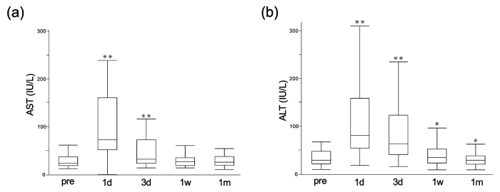
Fig. 1 Changes in transaminases in the perioperative period. (a) AST increased at 1 day and 3 days and improved at 1 week postoperatively. (b) The same trend is true for ALT, which increased at 1 day and 3 days and 1 week postoperatively. (*P < 0.01 **P < 0.001, Wilcoxon signed-rank test, compared to preoperative values) AST, aspartate aminotransferase; ALT, alanine aminotransferase.
In the long term, AST show significant changes (P = 0.0001, Friedman’s test). The mean values were 20[15–28]U/L at 3 months, 17[14–19]U/L at 6 months, and 17[14–20]U/L at 12 months postoperatively, showing a significant decrease compared to the preoperative values (P = 0.005, P < 0.001, P < 0.001, respectively, Wilcoxon signed-rank test) , with a decreasing trend (Fig. 2a). In the long term, the same significant changes can be seen for ALT (P < 0.0001, Friedman’s test). The mean values were 19[14–26]U/L at 3 months, 14[10–18]U/L at 6 months, and 14[12–23]U/L at 12 months postoperatively, showing a significant decrease compared to the preoperative values (P < 0.001, respectively, Wilcoxon signed-rank test) , with a decreasing trend (Fig. 2b).
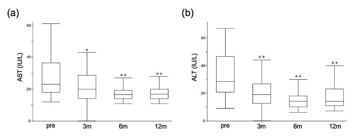
Fig. 2 Changes in transaminases up to 12 months after LSG. (a) In the mid-to-long-term postoperative follow-up, AST values decreased at 3, 6, and 12 months. (b) ALT values decreased at 3, 6, and 12 months postoperatively (. *P < 0.01 **P < 0.001, Wilcoxon signed-rank test, compared to preoperative values) LSG, laparoscopic sleeve gastrectomy.
After postoperative 12 months, BMI decreased from 42.6 kg/m2 to 32.1 kg/m2 (P < 0.0001, Wilcoxon signed-rank test). The total weight loss rate (%TWL) and the excess weight loss rate (%EWL) were 23.4%[19.3–30.9]and 59.5%[45.0–73.8], respectively, at 12 months postoperatively.
As for hepatobiliary enzymes other than AST ALT, T-Bil increased from 0.6 to 0.7mg/dL (P = 0.034, Wilcoxon signed-rank test) but there was no significant change in D-Bil (Pre: 0.1mg/dL, Post 0.1mg/ dL, P = 0.21, Wilcoxon signed-rank test). Regarding inflammatory reactions, CRP significantly decreased from 0.5 to 0.1mg/dL (P < 0.0001, Wilcoxon signedrank test). In lipid metabolism, HDL-C significantly increased from 50 to 57 mg/dL (P < 0.0001, Wilcoxon signed-rank test) while LDL-C significantly decreased from 122 to 113 mg/dL (P = 0.0003, Wilcoxon signedrank test) , and TG significantly decreased from 209 to 89 mg/dL (P = 0.0003, Wilcoxon signed-rank test). As for imaging findings, VS ratio decreased from 0.40 to 0.25 (P = 0.001, Wilcoxon signed-rank test).
The preoperative LS ratio was 0.87[0.69–1.12], and fatty liver was observed in 56.5% (26/46) of patients. However, in CT taken 12–24 months postoperatively, the LS ratio improved to 1.26[1.16– 1.34](P < 0.0001, Wilcoxon signed-rank test , Fig. 3a) and fatty liver decreased to 5.0% (1/20) (P < 0.001, χ2test) compared to preoperative values and numbers. As for liver fibrosis, no change was observed within the 12 months postoperative in the Fib-4 index (0.86[0.58–1.16]preoperative, and 0.90[0.74–1.10], 0.88[0.64–1.24], 0.92[0.68–1.12], and 0.96[0.67–1.22]at 1, 3, 6, and 12 months postoperatively, P = 0.15, Friedman’s test) (Fig. 3b).
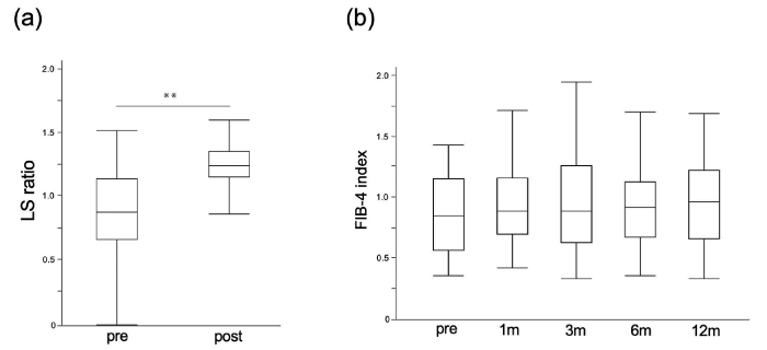
Fig. 3 The effect of LSG on liver fattening and fibrosis. (a) The preoperative LS ratio in plain CT was 0.87[0.69–1.12], and the LS ratio at 12–24 months after LSG significantly improved to 1.26[1.16–1.34]. (** P < 0.0001, Wilcoxon signed-rank test) (b) Fib-4 index did not change 1, 3, 6, and 12 months postoperatively. (P = 0.15, Friedman’s test) LS ratio, liver to spleen ratio; CT, computed tomography.
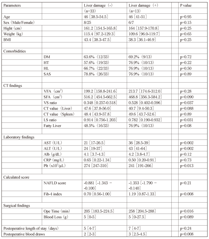
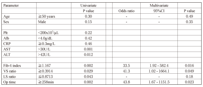
Differences in clinical factors were analyzed between groups with and without perioperative liver damage to identify factors associated with postoperative liver damage. No differences were found in age, sex, body size, or comorbidities, but statistically significant differences were found in VS ratio (P = 0.037, Mann–Whitney U test) and LS ratio (P = 0.031, Mann–Whitney U test) on CT, transaminases (P = 0.002, Mann–Whitney U test) and platelets (P = 0.013, Mann–Whitney U test) on blood tests, Fib-4 index (P = 0.008, Mann–Whitney U test) , and operative time (P = 0.016, Mann–Whitney U test) (Table 1).
Patients with perioperative liver damage stayed in the hospital for a median of 7 days longer than those without liver damage (5 days) , although this difference was not statistically significant (P = 0.24, Mann–Whitney U test). The number of postoperative blood tests was significantly higher in the group with hepatic impairment (3 vs. 2, P = 0.008, Mann–Whitney U test) (Table 1).
Table 1 Patient characteristics with or without perioperative liver damage
The cutoff values for LS ratio, VS ratio, Fib-4 index, and operative time, with perioperative liver damage as the outcome, were determined by ROC curves. LS ratio has an area under the curve (AUC) of 0.706 with a cutoff value of 0.8713; VS ratio was AUC of 0.699 with a cutoff value of 0.3914; operative time was AUC of 0.730 with a cutoff value of 258 min; Fib-4 index was AUC of 0.755 with a cutoff value of 1.167 (Fig. 4). The matrix scatter plot shows that each of these four continuous variables is uncorrelated as shown in Fig. 5.
The univariate and multivariate analyses using logistic regression analysis revealed the risk factor of perioperative liver damage, including high Fib-4 index (odds ratio[OR]= 33.5, 95% confidence interval[CI]: 1.92–582.4, P = 0.016) , high VS ratio (OR = 41.3, 95% CI: 1.02–1664.1, P = 0.049) , and long operative time (OR = 43.8, 95% CI: 1.67–1151.5, P = 0.023) (Table 2).
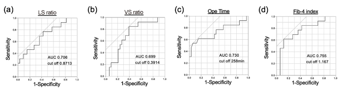
Fig. 4 ROC curves for setting the cutoff values for liver injury. ROC curve for (a) LS ratio, (b) VS ratio, (c) Operative time, and (d) Fib-4 index, with liver injury as an outcome.
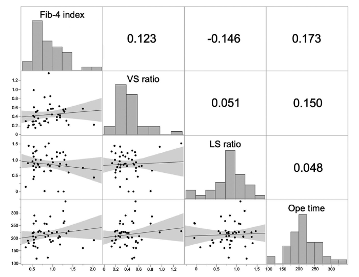
Fig. 5 Matrix scatter plot. Fib-4 index, VS ratio, LS ratio, and Operative time had no significant correlation with each other.
Table 2 Univariate and multivariate analysis of perioperative liver damage
A 42-year-old female patient had a preoperative BMI of 35 kg/m² and comorbidities of fatty liver, HTN, dyslipidemia, and SAS. After repeated rebounds from weight loss and inadequate medical treatment response, LSG was performed. Preoperative blood test results showed a slightly elevated inflammatory response and mildly elevated transaminases (Table 3). CT showed marked fatty liver and enlarged lateral zone, with a VS ratio of 0.63 and LS ratio of 0.78 (Fig. 6a). The operative time was 208 min, blood loss was 200 g, and the liver was elevated using the Nathanson River retractor (Fig. 6b).
Blood tests showed abnormally high transaminases (AST of 6,053 U/L, ALT of 3,650 U/L) and a markedly low platelet count of 26,000 1 day postoperatively.
A CT scan showed a contrast-impaired area in the outer hepatic area where the liver had been raised (Fig. 6c, d). The patient was diagnosed with thrombocytopenia and disseminated intravascular coagulation due to liver damage and splenic hyperfunction caused by intraoperative pressure drainage. The patient was conservatively treated with platelet transfusions, hepatoprotective drugs, and close follow-up blood tests. The patient was discharged 8 days postoperatively.
Table 3 Preoperative laboratory data
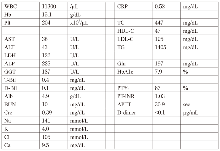
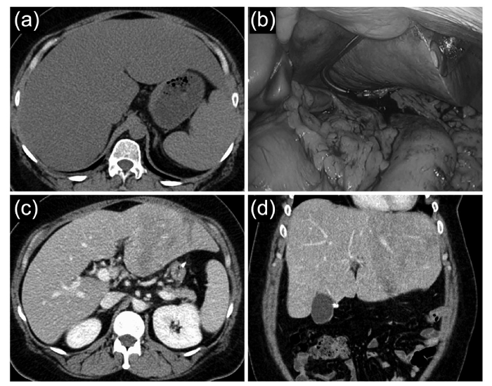
Fig. 6 Case presentation. (a) Preoperative plain CT showed marked fatty liver and enlarged lateral zone. (b) The liver was elevated using the Nathanson River retractor .(c, d) The enhanced CT scan showed areas of poor contrast in the outer hepatic zone.
This study revealed that high Fib-4 index, high VS ratio and long surgical duration are independent risk factors for perioperative liver damage for patients with obesity by the multivariate logistic regression analysis.
Perioperative liver damage is often a problem in epigastric surgery that requires liver elevation[6]. Additionally, the effect of CO2 on the decreased intestinal blood flow due to pneumoperitoneum is considered one of the causes of postoperative liver damage. Laparoscopic gastrectomy has been reported with more liver damage than open gastrectomy[7]. Although the above mechanisms suggest that patients with high BMI, high visceral fat ratio, and cirrhosis, which require more effort to expand the field of view by liver elevation, and long procedure time, which is suggested to be associated with prolonged hepatic ischemia time, are thought to be risks for postoperative liver damage, there are no reports that have clarified this. Previous reports examining risk factors for elevated liver enzymes after laparoscopic gastrectomy have reported the use of Nathanson River retractors and dissection of the accessory left hepatic artery[8], but reports on the risk of perioperative liver injury, including LSG, are very limited.
In the field of bariatric or metabolic surgeries, raising the liver to exposure the gastroesophageal junction and the angle of His when processing the cardia is necessary. Additionally, patients often have a thick abdominal wall and an enlarged liver, which makes liver elevation and surgery difficult. Many cases have preoperative liver dysfunction due to obesity[9,10]. The NAFLD frequency in the health examination of Japanese adult subjects is 30%–40% in males and 10%–20% in females. The prevalence of NASH is estimated as 2%–8% among adults since 10%–20% of NAFL is reported as NASH[11]. The prevalence of NAFLD by sex and age directly reflects the frequency of obesity. Recently, MAFLD is considered more appropriate umbrella term[1]. LSG in children with obesity induces an improved MAFLD-related metabolic derangement and liver damage, and more and more cases and indications are expected to expand in the future[3,12]. Therefore, various surgeons have devised new techniques for liver retraction to overcome this problem[13,14]. The surgical number of patients with morbid obesity with MAFLD is expected to increase in the future; thus, an accurate preoperative risk assessment is desirable.
Since the Fib-4 index is calculated mainly from transaminases, it is not hard to imagine that a high Fib-4 index can predict perioperative liver damage. However, independent of the Fib-4 index, VS ratio and operative time were found to be risk factors. Most postoperative liver disorders are transient; however, a risk assessment is particularly important in surgeries for obesity because they may cause liver disorders that require platelet transfusion, as in the presented case, and may lead to longer hospital stays and more frequent blood tests. Intervention to decrease VS ratio may be possible as preoperative management. The visceral fat is generally known to decrease early in weight loss; thus, reducing the VS ratio due to preoperative weight loss may avoid liver damage. With a high risk of liver damage, shortening the operative time must be decided as much as possible.
As a study limitation, the biopsy required for NAFL or NASH evaluation is not performed at this facility. Liver steatosis is graded based on the percentage of fat within the hepatocytes and the average LS ratio corresponding to steatosis grade 1 (mild, 5%–33%) is said to be 0.88 ± 0.28[15,16]. Generally, an LS ratio of 0.9 is used as the cutoff value for fatty liver diagnosis, and the patient is considered without fatty liver with an LS ratio of >1.1[17,18]. From the above study reports, CT was used as a surrogate for assessing hepatic lapidification. In addition, liver fibrosis is also evaluated only by Fib-4 index. Of course, evaluation of fibrosis by transient elastography (FibroScanⓇ) , or blood markers such as Mac-2 binding protein glycosylation isomer (M2BPGi) would be desirable, but this has not been done in this study due to lack of equipment. Recent studies have shown that FIB-4 has poor performance in young or obese diabetic patients[19], and we believe it is necessary to evaluate fibrosis with multiple modalities in the future.
The outcomes of this work suggest that LSG can improve liver fatness at least for 1–2 years postoperatively; however, no improvement was observed in fibrosis (Fib-4 index) in the same period. In NAFLD, 5%–8% will develop cirrhosis within 7–21 years, and 30%–50% will develop liver fibrosis within 3–10 years in NASH. The result is thought as a prognosis worsening for life[20]. It takes a certain period for fatty liver to develop into steatohepatitis and fibrosis, and the period studied in this study is considered insufficient to determine the effect. Additionally, the present study did not include cases with advanced fibrosis; thus, future surgical cases with more fibrosis should be included in the analysis.
YM, MK, HH, HS, RO and HM involved in surgical treatment. HO and KY involved in the medical treatment of the patient. Statistical analyses were performed by YM, using the image data values extracted by KH and YK. The manuscript was written by YM under the supervision of HO, YK, HH and HM.
There is no financial support associated with this report.
H. M. is a member of the Editorial Board of the Chiba Medical Journal.
This study was approved by the ethical committee of Chiba University Graduate School of Medicine. (assignment number 3043) Consent for this study in 46 cases was given on an opt-out basis. In addition, consent for the case report was obtained verbally.
The data that support the findings of this study are available from the corresponding author, YM, upon reasonable request.
We would like to thank Enago (www.enago.jp) for English language editing.
Address correspondence to Dr. Yasunori Matsumoto.
Department of Frontier Surgery, Chiba University Graduate
School of Medicine, Chiba University, 1-8-1 Inohana, Chuou-Ku,
Chiba 260-8670, Japan.
Phone: +81-43-226-2109.
E-mail: ymatsumoto@chiba-u.jp
