


Chiba Medical J. 99E:51-56,2023
doi:10.20776/S03035476-99E-5-P51
〔 Case Report 〕
Marina Sakata1), Yuri Kawashima2,3), Akihiko Adachi1)*
Kosuke Adachi1), Yuya Furukawa1), Tomoko Yoneyama-Sarnecky1)
and Michio Nakamura1)
1) Department of Neurosurgery, Japanese Red Cross Narita Hospital, Narita 286-8523.
2) Department of General Internal Medicine, Japanese Red Cross Narita Hospital, Narita 286-8523.
3) Department of Life Sciences, Graduate School of Arts and Sciences, The University of Tokyo, Tokyo 153-8902. *Contact information for the corresponding author.
(Received March 24, 2023, Accepted March 27, 2023, Published December 10, 2023.)
Displacement of the tip of an implanted tube is a serious complication in medical practice. Herein, we report two cases of unintended advancement of a drainage catheter during external ventricular drainage (EVD). Fortuitously, the patient did not present with any associated symptoms. The similarities between the two patients included the technique used, such that the EVD catheter was inserted through the occipital area, as well as the patient’s postoperative tendency to be restless and rub the back of his/her head against a pillow. In other words, the same phenomenon occurred in the postoperative period of two surgeries performed at different times over an interval of two years by different surgical teams. Therefore, it is considered appropriate to investigate the constitutive causes from the viewpoint of medical safety. Our detailed material science and mathematical considerations, especially topological considerations, proved that the outer diameter of the catheter decreases with tension and twisting. Therefore, strategies to prevent catheter migration include the following. 1) EVD should be tunneled subcutaneously to an appropriate length, and the EVD exit site should be adequately far from the main incision site. 2) The distal end of the ligature thread on the distal drainage catheter might be sutured to the skin instead of being cut. 3) A purse-string suture at the catheter exit site and modified Roman sandal tie could be created. 4) Frequent rubbing against the pillow or bed at the exit site of the EVD catheter should be avoided.
external ventricular drain, proximal intraventricular migration, complication, silicone tube, hydrocephalus
Unintended displacement or straying of tubes can occasionally occur during medical interventions and is reported previously [1,2]. Possible causes can be categorized as follows: factors on the part of the healthcare provider, such as inaccurate techniques; the nature of the device, such as fragmentation; performance of the technique itself, such as disconnection or theoretically unavoidable loosening between the tube and the thread; and factors on the part of the patient, such as self-removal.
We report on our experience of two cases with ventricular catheter straying.
External ventricular drainage (EVD; Silascon TM Ventricular Drainage, Kaneka Medical Products, Osaka, Japan) was inserted through the posterior horn in the occipital region in both patients. EVD catheters were fixed to the scalp at the puncture site using a 2-0 silk thread. The catheters were ligated at the proximal and distal parts (a few centimeters from the proximal ligation site) for anchoring (Fig. 1b, 3b and 6g). The catheter was connected to a drainage bag via a ventricular drainage circuit to provide a completely closed system for EVD. The catheter was directed into the lateral ventricle, 8 cm from the outer plate of the skull, through the brain parenchyma. Computed tomography (CT) and magnetic resonance imaging (MRI) were performed to evaluate the postoperative hemorrhage and the position and trajectory of the EVD catheter.
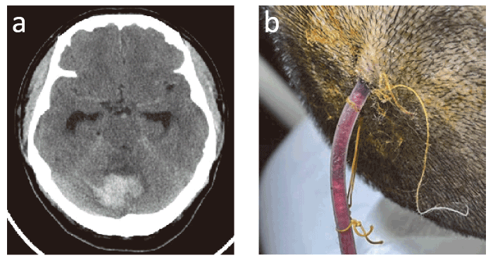
Fig.1 Case 1. (a) CT scan of the cerebellar hemorrhage; (b) Catheter exit site immediately before the drainage catheter was removed. Note that the white lint is a piece of gauze and not an unraveled silk thread. CT, Computed tomography.
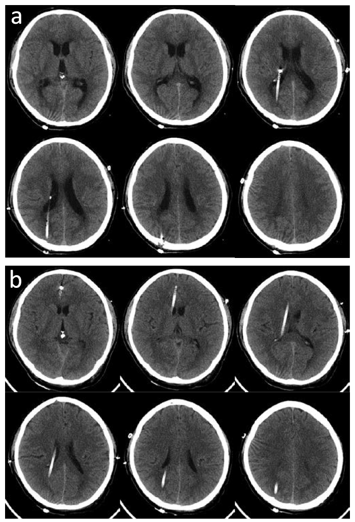
Fig.2 CT scans of Case 1. (a) Immediately after surgery; (b) 5 days after surgery.
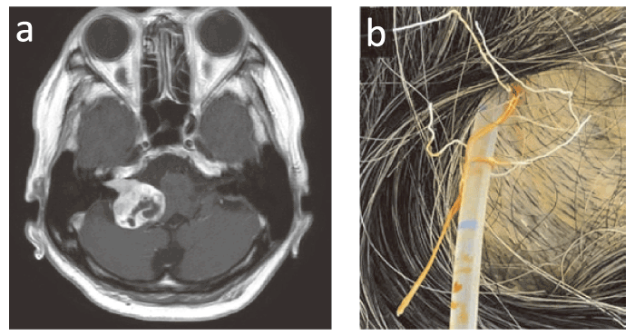
Fig.3 Case 2. (a) Contrast-enhanced MRI of vestibular schwannoma; (b) Catheter exit site just before the drainage catheter was removed. Note that the white lint is a piece of gauze and not an unraveled silk thread. MRI, magnetic resonance imaging.
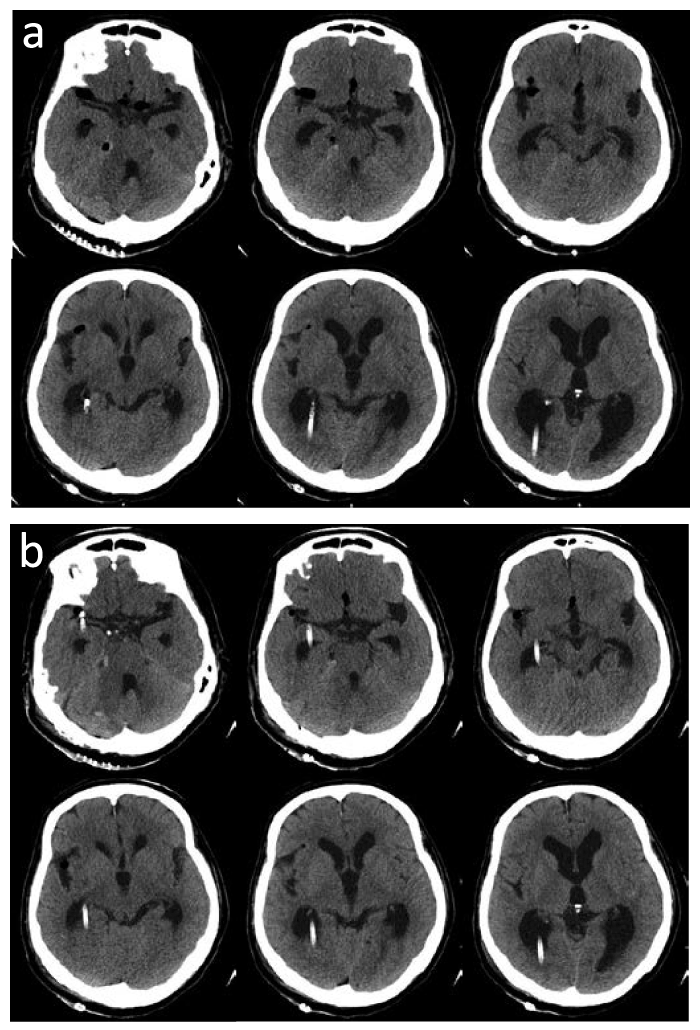
Fig.4 CT scans of Case 2. (a) The day after surgery; (b) Two days after surgery
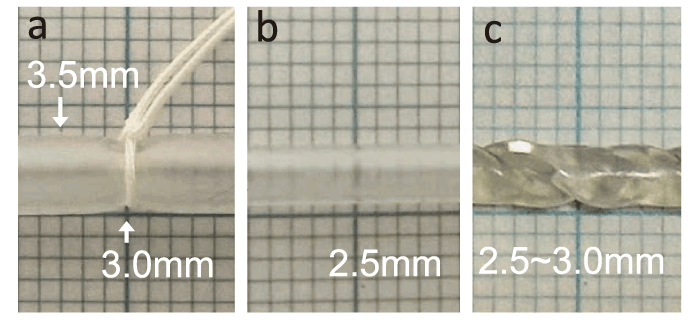
Fig.5 Ventricular catheters in various states due to external forces. Note the changes in outer diameter due to compression, stretching, or topological change, by torsion. (a) Ligated with 2-0 silk thread; (b) Stretched; (c) Twisted.
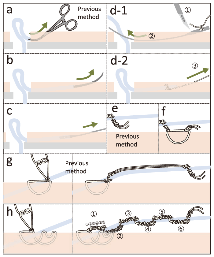
Fig.6 Schemas of possible strategies for preventing catheter migration. (a, e. g) Previous method; (b, c, d) Long subcutaneous tunnel; (f) Ligation to the skin in two positions; (h) Purse-string suture at the catheter exit site and a modified Roman sandal tie.
A 50-year-old man presented with disturbance of consciousness due to acute hydrocephalus associated with cerebellar hemorrhage (Fig. 1a) and was treated with EVD along with decompression craniectomy to remove the hematoma. His consciousness status improved immediately after surgery. Compared with the CT images immediately after surgery (Fig. 2a), the images on postoperative day 5 (Fig. 2b) showed migration of the catheter into the right frontal lobe. The drainage volume from the EVD catheter was low; therefore, the catheter was removed on the fifth postoperative day. As the hydrocephalus had already improved with the removal of the hematoma, there were no symptoms associated with catheter removal. A photograph of the drainage catheter immediately before its removal is shown in Figure 1b.
A 66-year-old woman with hydrocephalus associated with vestibular schwannoma (Fig. 3a) underwent EVD during craniotomy for tumor removal. Compared with the CT images on the day after surgery (Fig. 4a), the images on the second postoperative day (Fig. 4b) showed migration of the catheter into the tip of the right temporal lobe. The catheter was removed on the second postoperative day. A photograph of the catheter taken immediately before its removal is shown in Figure 3b. A ventriculoperitoneal shunt was inserted for hydrocephalus one month after the surgery, and symptoms such as dizziness and urinary incontinence disappeared.
In both cases, catheter migration was observed a few days after the surgery. Here, we discuss the causes of this complication.
First, we evaluated the surgeon factors related to catheter migration. In both cases, catheter placement, skin suturing, and drain ligation were performed by different surgeons. The surgery in case 1 was performed by a neurosurgeon who was 10, 14, and 34 years postgraduation. In case 2, the patient was operated on by a neurosurgeon 4, 26, and 36 years post-graduation. Therefore, this cannot be simply attributed to surgeonrelated factors. No significant suture loosening was observed (Fig. 1b and 3b). If the thread is ligated at an angle to the catheter, it moves and loosens [3]. However, Figure 1b and 3b show that the threads were ligated perpendicular to the catheter.
Second, we estimated the nature of the device and the perfection of the procedure. The deformation of the catheter was initially examined. The outer diameter of the catheter essentially changes owing to external factors, such as ligating (Fig. 5a), stretching (Fig. 5b), and twisting (Fig. 5c). It is also possible that stretching and twisting reduce the outer diameter more than ligation. This may have created a gap between the ligature thread and drainage catheter, causing the catheter to migrate. The so-called “ladder” method (Fig. 1b, 3b and 6g), in which the catheter is ligated at two points, creates friction at both points in the direction the catheter is pulled out. However, only the proximal ligature works to prevent advancement, while theoretically the distal ligature does not. Moreover, subcutaneous tunnels were created through which the catheters were passed by incising the skin with a sharp scalpel and pulling out the catheters with hemostatic forceps (Fig. 6a). However, the subcutaneous tunnels that can be created using this method are short, and friction in the subcutaneous tunnel area is considered insufficient.
Finally, we suggest several measures for preventing unintended catheter advancement. It is necessary to recognize that the diameter of the catheter can be reduced by external forces, so that the friction between the catheter and the thread can decrease, even if the catheter seems to be tightly bound to the thread. To prevent the catheter from moving, the following methods can be considered:
1) Instead of pulling the catheter out using hemostatic forceps (Fig. 6a), use a long metal needle, i.e., trocar (Fig. 6b) or passer (Fig. 6c) firmly inserted into the distal end of the catheter, or use a passer and silk thread (Fig. 6d-1 and 6d-2), to pass the catheter under the skin and create a long subcutaneous tunnel.
2) Suture a thread at the distal knot to the skin (Fig. 6f), instead of cutting it immediately after the distal knot (Fig. 6e).
3) Create a purse-string suture at the catheter exit site and create a modified Roman sandal tie [3], a combination of six knots along the drain and one loop around the drain, repeated six times (Fig. 6h).
4) Avoid frequent rubbing against the pillow or bed at the exit site of the EVD catheter.
M. S. wrote the manuscript; Y. K. was involved in the discussion and provided Figures 5 and 6; A. A. initiated and directed all aspects of the project; Y. F., A. A., and M. N. performed the surgery in case 1; and K. A., T. YS. and M. N. performed the surgery in case 2. All the authors read and approved the final version of the manuscript.
This report received no specific grants from any funding agencies in the public, commercial, or not-forprofit sectors.
All authors declared no competing interests including (but not limited to) employment, honoraria, stocks, patents, materials, and equipment.
This report complies with all relevant national regulations and institutional policies. Furthermore, it is in accordance with the tenets of the Helsinki Declaration. Patient consent for the release of patient information was obtained, which can be provided upon request.
The datasets generated during the current study are available from the corresponding author on reasonable request.
Address correspondence to Dr. Akihiko Adachi.
Department of Neurosurgery, Japanese Red Cross Narita Hospital,
Iida 90-1, Narita 286-8523, Japan.
Phone: +81-90-5515-2741.
Fax: +81-43-226-2159.
E-mail: adachi-cib@umin.ac.jp
