


Volume 88, Number 1
doi:10.20776/S03035476-88E-1-P1
[Original Paper]
Koki Yamashita*, Yasufumi Kasagi1) Ken Nakazawa2) and Ichiro Shimoyama1)
(Received July 12, 2011, Accepted November 8, 2011)
The effect of delayed auditory feedback on the activity of the bilateral prefrontal region of the brain was investigated by functional near-infrared light spectroscopy (fNIRS). Ten volunteers read aloud a story written in Japanese and alternatively listened to their self-voice through inner ear earphones with no delay or a designated 200-ms delay, repeating the task five times each. The changes in total hemoglobin(Hb) and oxygenated hemoglobin(Oxy-Hb) levels of the prefrontal region and in cutaneous total Hb levels over the mid forehead were monitored. Duration of reading time was also recorded. The mean duration in reading time with delayed feedback was significantly prolonged compared to without the delay. The reading time of the first delayed feedback was significantly prolonged. There was no correlation between cutaneous and cerebral total Hb levels; therefore, fNIRS data were most likely not affected by cutaneous blood flow. Changes in Oxy-Hb levels between delayed and non-delayed feedback of self-voice were not detected in this study. Increase of Oxy-Hb levels in the left prefrontal cortex was observed with and without delayed auditory feedback most likely due to the linguistic-related functional differences between left and right hemispheres of the brain.
delayed auditory feedback, oxygenated hemoglobin, prefrontal cortex, functional near-infrared spectroscopy(fNIRS)
Auditory feedback is important in detecting and correcting errors during speech. The human brain detects differences between intended sounds and performed output and uses them to alter motor patterns so that proper sound production is eventually achieved[1]. Temporal asynchrony between speech production and its feedback such as delayed auditory feedback causes the speaker to speak less fluently[2,3]. The delayed auditory feedback effect is observed as articulator changes, including slower speech[4]. Recent research in behavioral measurement has shown the maximal disruption to occur with a delay of approximately 200 ms[5]. While speech fluency is maintained without any conscious effort under real-time auditory feedback, conscious self-monitoring for overt-speech processing is required under delayed auditory feedback. The array of brain imaging study techniques now available may help in understanding the differences in brain activity under real-time auditory and delayed auditory feedback.
A major aim of functional mapping of the human brain is to visualize internal operations occurring for various brain functions. Functional brain mapping can now be achieved with the establishment of modern neuro-imaging techniques such as positron emission tomography (PET) and functional magnetic resonance imaging (fMRI). Alternatively, near-infrared spectroscopy (NIRS) is a relatively novel noninvasive optical technique to measure changes in oxygenated hemoglobin (Oxy-Hb) and deoxygenated hemoglobin concentrations in the cerebral cortex. The methodology of NIRS was first proposed by Jöbsis in 1977[6], and its technique was initially designed for clinical monitoring of tissue oxygenation[7-9].
In the 1990s, this technique was utilized in functional neuro-imaging studies, and referred to as functional near infrared spectroscopy (fNIRS)[10-12]. Brain activation is accompanied by a complex sequence of cellular, metabolic, and vascular processes. The relationship between local neural activity and subsequent changes in cerebral blood flow, called neurovascular coupling, is considered an index of neural activity, rather than showing concrete activity of the brain. Like fMRI and PET, fNIRS can detect brain activity using this method of neurovascular coupling. fNIRS is considered to be a useful tool for neuro-imaging studies because it is completely non-invasive and does not require strict motion restriction compared to PET and fMRI. With such advantages, fNIRS has been used to investigate motor and cognitive brain activities, including visual[13-15], auditory[16], speech[17], and motor movements[18]. In the last decade, fNIRS has expanded its applications to developmental psychology[19,20]and psychiatry[21-29]. As fNIRS is a relatively novel and still not fully developed tool to investigate brain activities, further research on the accuracy and reliability of fNIRS is warranted. One concern arises from the unclearness of the selective detection of fNIRS signals arising from the cerebral tissue. The subcutaneous blood flow may influence the near-infrared light as the input sensor of fNIRS can detect the near-infrared light carrying the information on not only cerebral tissue but also extracerebral tissue.
In this study, fNIRS was applied to examine the effect of delayed auditory feedback on the activity of the bilateral prefrontal region of the brain. We considered that a delayed auditory feedback task is associated with executive function, as well as working memory, a neural mechanism that provides and supports temporary storage and manipulation of information in the service of cognition. We focused on bilateral prefrontal brain activity during the delayed auditory feedback task with a delay of 200 ms. Hemoglobin changes in the subcutaneous area were monitored to determine the influence of the subcutaneous blood flow on fNIRS data.
The study was conducted in a quiet room located at the Center for Frontier Medical Engineering, Chiba University. Ten healthy volunteers (9 male and 1 female, aged 19 to 60 [mean 41.1±16.1]), who were right-handed and whose first language was Japanese, participated in this study. All participants confirmed to have no history of auditory problems. Written informed consent was obtained from each participant. Participants read aloud one of Aesop’s Fables (in Japanese) as they listened to their own voices through inner ear earphones. A representative short story, the Japanese version of one of Aesop’s Fables, is shown in Figure 1(Fig. 1) . All participants confirmed that they had never read the story before. The text of the story was displayed on paper in front of the participants.

Fig. 1
The Aesop’s fable read aloud by volunteers (The Stomach and the Feet)
Before reading, each participant sat upright in a chair with a headrest. To minimize movement artifacts caused by head and body movement during monitoring, participants set their heads on the headrest and were instructed not to move. They were instructed to relax before starting the first task. At a given auditory cue to start the task, they started reading the story. During the study, they read the story aloud and alternatively listened to their self-voice in two phases, without delay (non-delay) or with a 200-ms delay (delay) for a total of five times each. Each task was initiated by the given auditory cue.
Self-voice was controlled by an auditory amplifier and a sound delay system (real-time processor, RM2, TDT, USA). Total Hb and Oxy-Hb levels in the bilateral prefrontal region of the brain were monitored every 1/6 s using two-channel fNIRS (NIRO-200, Hamamatsu, Japan). Probes of the fNIRS were set at Fp1 and Fp2 sites, above the eyebrows and sinuses. Changes in total Hb of the subcutaneous blood flow were monitored over the mid forehead at 10 Hz (CDF-2000, CyberMed, NEC Infrontia, Japan) (Fig. 2). During the task, each participant’s voice was recorded with a voice recorder at 22.1 kHz to examine the duration of reading time.
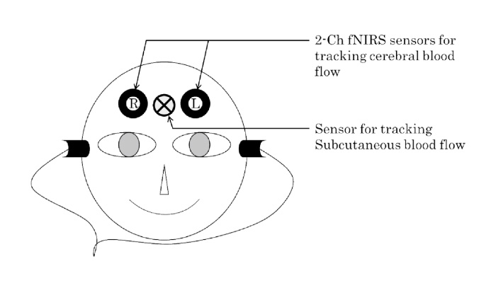
Fig. 2
Sensors on the forehead
Total Hb and Oxy-Hb changes in the bilateral prefrontal cortex were monitored during each non-delay and delay phase, as well as during pre-reading as a reference for physiological fluctuation of the restful state. The relationship between the subcutaneous total Hb and bilateral prefrontal cortical total Hb changes, as well as the duration of reading time, was assessed. The relative changes of Oxy-Hb levels were monitored from the auditory cue (set as 0) to the point the participant finished reading the story, and the results of each task were averaged. The changes in Oxy-Hb levels detected by fNIRS and the duration of the average reading time were analyzed using ANOVA and Tukey-Kramer HSD. Probability values of <0.05 were considered to be statistically significant. The correlation of the time-course changes in total Hb between the bilateral prefrontal cortex of the brain and subcutaneous blood flow was examined using Pearson’s correlation coefficient.
The mean duration in reading time with delayed feedback was 26.5±3.9 s, which was significantly prolonged compared to 23.14±3.6 s without the delay (P<0.001) (Fig. 3). The mean duration in reading time with delayed feedback was also significantly prolonged at the first reading (P<0.05); however, the difference was not significant within each repeated reading set (Fig. 4).
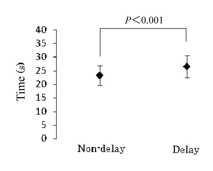
Fig. 3
Duration of reading time (all data included)
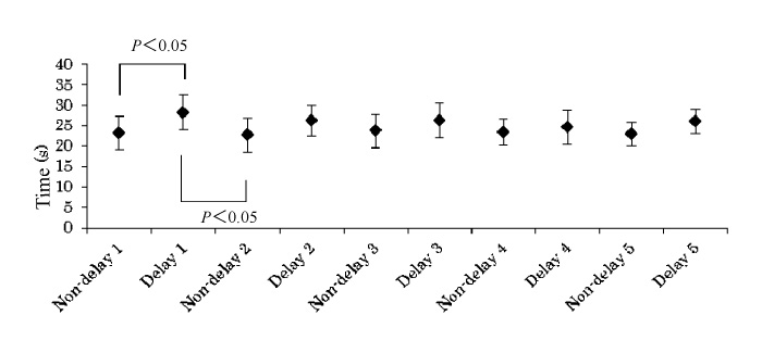
Fig. 4
Chronological changes in duration of reading time
The mean correlation coefficient values between cutaneous and left prefrontal cortex, cutaneous and right prefrontal cortex total Hb changes were 0.03±0.2 and 0.06±0.1, respectively. The mean Oxy-Hb levels of the left and right prefrontal cortices were -7.35±3.6 and -0.9±2.7 in the pre-reading phase; 0.72±0.9 and 1.7±0.7 in the non-delay phase; and 0.37±0.9 and 1.13±0.7 in the delay phase, respectively. There were no significant differences in Oxy-Hb levels between the left and right prefrontal cortices in each phase. Compared to the pre-reading phase, Oxy-Hb levels of both the left and right prefrontal cortices were increased in the non-delay and delay phases. Although there was no significant difference in the right prefrontal cortex, there was a significant difference between the pre-reading and non-delay phases (P<0.05), as well as between the pre-reading and delay phases (P<0.05), in the left prefrontal cortex (Fig. 5) . Notably, Oxy-Hb levels varied within each repeated reading set. Moreover, differences in Oxy-Hb levels of both the left and right prefrontal cortices were not significant between non-delay and delay phases.
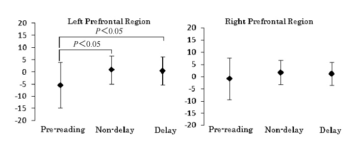
Fig. 5
Oxy-Hb levels on the left and right prefrontal regions (all data included)
The effect of delayed auditory feedback on the activity of the bilateral prefrontal cortex of the brain was investigated using 2-ch fNIRS. fNIRS is a useful tool to investigate brain activities during auditory stimulation as this instrument runs without making any noise which may influence readings of auditory brain activities[30]. Therefore, fNIRS is considered a preferable technique for this and other auditory studies.
In this study, the mean reading time for completion of the story was significantly prolonged when self-voice was delayed compared to when it was not, which confirms that the disruption successfully occurred with delayed feedback by the designated time (200 ms). This significant increase in reading time was particularly seen at the first reading. With each subsequent reading, however, the difference in reading times with and without delayed feedback disappears, most likely due to the learning effect.
To investigate the influences on subcutaneous blood flow during the auditory task, we assessed the relationship between changes in subcutaneous total Hb and bilateral prefrontal cortical total Hb. No correlations were observed between subcutaneous and left prefrontal cortex total Hb changes and between subcutaneous and right prefrontal cortex total Hb changes. Thus, fNIRS data were most likely not affected by subcutaneous blood flow in this study. These results are limited in the fact that the corrected data of subcutaneous total Hb levels and bilateral prefrontal cortical total Hb levels were not from exactly the same area, and only total Hb levels were monitored and investigated.
Oxy-Hb levels of the left and right prefrontal cortices were decreased during a restful state before the reading task with no significant difference between the cortices. In contrast, the left prefrontal cortex was activated with significantly increases in Oxy-Hb levels when individuals read a story aloud with or without delayed feedback. These results suggest a relationship of the left prefrontal cortex with reading and listening to self-voice. In general, the left hemisphere is involved in controlling language related movement, whereas the right hemisphere plays a role in non-verbal abilities. Therefore, significant increases in Oxy-Hb levels in left prefrontal cortex during the reading the story observed in this study might be due to the functional differences related to linguistics between the left and right hemispheres of the brain.
The changes in Oxy-Hb levels have a high association with a change in regional cerebral blood flow levels, and the area showing increasing regional cerebral blood flow reflects the increase in nerve activity in the area[31]. Therefore, the area showing increasing Oxy-Hb levels is considered to have increased nerve activity. We speculate that the changes in Oxy-Hb levels observed in this study are related to regional brain activities. First of all, this study did not show a significant increase in Oxy-Hb levels during the task compared to the restful state in the right prefrontal cortex, which might suggest that the task did not activate the right prefrontal cortex, as this task requires the linguistic process of the brain. On the other hand, a decrease in Oxy-Hb levels does not necessarily correspond to lack of nerve activities in that area. That is, an increase in Oxy-Hb levels may not be detected when there is more significant brain activity in the area outside the monitored area. This surrounding area would thus require a greater volume of Oxy-Hb and may not lead to an increase in per se the monitored area. Secondly, for the left prefrontal cortex, the Oxy-Hb levels significantly increased during the task compared to during the restful state, correlating to significant brain activity during the task. However, the Oxy-Hb levels were not significantly different between tasks with and without the delay. During the task, a high volume of Oxy-Hb had most likely moved into the cortex thereby limiting the capacity for increased blood flow.
In this study, we focused on executive function and working memory. We investigated the Oxy-Hb changes in the frontal cortex, although a delayed auditory feedback task may influence brain activities in regions of the brain that are linked to speech production, including Broca’s area and Wernicke area. A brain mapping study of delayed auditory feedback using fMRI suggested that tempo-parietal regions function as a conscious self-monitoring system to support an automatic speech production system[32]. Moreover, recent findings suggest that the cerebellum plays a role in a range of cognitive functions, as well as learning, working memory, and executive functions[33,34]. Therefore, monitoring the changes in Oxy-Hb levels by multi-channel fNIRS appears suitable for the detection of differences in brain activities in regions of the brain other than the frontal cortex, including the tempo-parietal regions and cerebellum. Studies have yet to investigate the brain activities of tempo-parietal regions and cerebellum under real-time auditory and delayed auditory feedback using fNIRS.
fNIRS is a useful tool for detecting blood flow in the cerebral cortex, although interpretation of the detected signal is still under investigation[35]. The uncertainty of which region in the brain is sampled by the near-infrared light and the complicated brain activities and blood flow of cerebral tissue has limited the acceptance of fNIRS[36,37]. Moreover, no method has been established for data analysis of fNIRS, such as statistical parametric mapping[38]which is used for fMRI and PET data analysis. fNIRS is still a novel neuroimaging tool, and its combination with other tools such as electroencephalogram or fMRI should be considered for obtaining more accurate and reliable data.
In this study, disruption of fluent speech as well as the significant increase of Oxy-Hb levels in the left prefrontal region was observed by 200-ms delayed auditory feedback. The significant increase of Oxy-Hb levels in the bilateral prefrontal during delayed auditory feedback was not detected by fNIRS. However, increases of Oxy-Hb levels in the left prefrontal cortex, but not right prefrontal cortex, were observed with and without delayed auditory feedback, most likely due to the linguistic-related functional differences between the left and right hemispheres of the brain. Multi-channel fNIRS has potential in monitoring the changes in Oxy-Hb levels for the detection of differences in brain activities in regions of the brain, in addition to the frontal cortex.
*Human Neurophysiology, Graduate School of Medicine, Chiba University, Chiba 263-8522.
1) Human Neurophysiology, Center for Frontier Medical Engineering, Chiba University, Chiba 263-8522
2) Department of Cognitive Behavioral Physiology, Graduate School of Medicine, Chiba University, Chiba 260-8670.
Tel.+81-43-290-3118. Fax. +81-43-290-3118. E-mail: kokiyamashita@hotmail.com
