


Chiba Medical J. 88E:41~46,2012
doi:10.20776/S03035476-88E-4-P41
[Original Paper]
Seiji Ohtori, Masaomi Yamashita, Yasuaki Murata, Yawara Eguchi
Yasuchika Aoki, Hiromi Ataka, Jiro Hirayama, Tomoyuki Ozawa
Tatsuo Morinaga, Hajime Arai, Masaya Mimura, Hiroto Kamoda
Sumihisa Orita, Masayuki Miyagi, Tomohiro Miyashita, Yuzuru Okamoto
Tetsuhiro Ishikawa, Hiroaki Sameda, Tomoaki Kinoshita, Eiji Hanaoka
Miyako Suzuki, Munetaka Suzuki, Takato Aihara, Toshinori Ito
Gen Inoue, Masatsune Yamagata, Tomoaki Toyone and Kazuhisa Takahashi
Chiba Low Back Pain Forum.
Department of Orthopedic Surgery, Graduate School of Medicine, Chiba University, Chiba 260-8670.
(Received January 5, 2012, Accepted January 27, 2012)
Purpose: Compression of the spinal nerve roots by lumbar spinal stenosis is a major clinical problem associated with intermittent claudication, pain and numbness. The pathological mechanism is reduced blood flow in nerve roots and degeneration of nerve roots. Prostaglandin E1(PGE1) is used for patients with peripheral arterial disease(PAD) to increase capillary flow around the main artery and improve symptoms; however, the ankle-brachial index(ABI) does not change after treatment.Lumbar spinal nerve roots contain somatosensory, somatomotor, and unmyelinated autonomic nerves, and improved blood flow in these spinal nerve roots may improve the function of nerve fibers innervating muscle, capillary, and main vessels in the lower leg, resulting in an increased ABI. The purpose of the study was to examine the hypothesis that limaprost, a PGE1 analog, improves blood flow in compressed and degenerated spinal nerve roots, leading to improved function of sympathetic and parasympathetic nerves in spinal nerve roots innervating the lower leg and a resultant increase in the ABI.
Methods: Limaprost(15 μg 3 times a day for 3 months) was administered orally to 49 patients with lumbar spinal stenosis. Low back pain and leg pain scores, walking distance, and ABI were measured before treatment and after 3 months of limaprost treatment.
Results: Low back pain, leg pain, and maximum walking distance significantly improved after limaprost treatment(P<0.05). ABI was also significantly increased by limaprost treatment(P=0.003).
Conclusions: This is the first investigation of changes in ABI in patients with lumbar spinal stenosis after treatment with a PGE1 analog. Our findings suggest that it is important to consider the role of the autonomic nervous system in spinal nerve roots associated with blood supply to the lower leg in patients with lumbar spinal stenosis.
Prostaglandin E1, Limaprost, Pain, Radiculopathy, Ankle brachial pressure index, Spinal stenosis
Compression of spinal nerve roots by lumbar spinal stenosis(LSS) is a major clinical problem associated with intermittent claudication, pain, numbness, and lack of normal sensitivity[1,2]. Such compression has been shown to induce neurophysiologic dysfunction, degeneration, and reduced blood flow in nerve roots in animal models and in humans[1,2]. Drug therapy is often used in patients with mild to moderate symptoms, mainly with administration of nonsteroidal antiinflammatory drugs(NSAIDs)[3,4].
Reduced blood flow in nerve roots induces neurogenic intermittent claudication, and drugs for improvement of blood flow in nerve roots have been developed. Prostaglandin E1(PGE1) causes vasodilation in both arterioles and venules[5,6]. In a clinical study, intravenous lipo-PGE1 administered for 10 consecutive days to 40 patients produced symptomatic improvement for a limited period in treatment of neurogenic intermittent claudication associated with lumbar spinal stenosis[7]. An investigation of 25 cases of lumbar spinal stenosis by myeloscopy revealed that the diameters of blood vessels in the cauda equina differed significantly from those in a control group[8]. In a separate study, myeloscopy also showed elimination of morphological changes in vessels along the cauda equina after administration of lipo- PGE1 in patients who originally had vessel dilation on the surface of the cauda equina[9]. These human studies show that PGE1 improved the microcirculation in spinal nerve roots and relieved symptoms in patients with lumbar spinal stenosis.
Patients with peripheral arterial disease(PAD) may be asymptomatic or present with a spectrum of symptoms including atypical leg pain, classic claudication, rest pain, and critical limb ischemia with gangrene[10]. PAD is diagnosed by assessing the ankle-brachial blood pressure index(ABI), a rapid and simple non-invasive diagnostic technique[10]. Exercise and drugs such as PGE1, cilostazol(a selective inhibitor of phosphodiesterase III and an antiplatelet drug), and angiotensin II type 1(AT1) receptor blockers improve the symptoms of PAD[10]. In general, these drugs increase flow in capillary vessels and do not effect the main arterial tract. Thus, most reports have shown that these therapies improve symptoms, but do not influence ABI[11,12].
The lumbar spinal ventral nerve roots and dura mater include somatic and autonomic nerves such as unmyelinated sympathetic and parasympathetic nerves that innervate the lower leg[13-15]. Therefore, we hypothesized that treatment with limaprost, a PGE1 analog, would increase blood flow on the surface of compressed and damaged spinal nerves, and that this would improve the function of somatosensory, motor and autonomic nerves innervating the main artery in the lower leg and lead to an increase in ABI. Improvement of blood flow in the lower leg after therapy for lumbar spinal stenosis has not been examined previously. Therefore, the purpose of the current study was to investigate the effect of limaprost on symptoms of lumbar spinal stenosis and on ABI.
The ethics committee of our institution approved the protocol for the human procedures used in this study. Informed written consent was obtained from each subject.
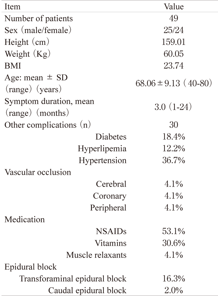
Patients The subjects were patients with low back and leg pain that had continued for at least 1 month. Patients who had previously undergone spinal surgery were excluded from the study. We also excluded patients with spinal tumor, infection, or trauma. Lumbar spinal stenosis was diagnosed on X-ray and magnetic resonance imaging(MRI) and by physical examination by spine surgeons. On MRI, the degree of spinal stenosis varied from slight to severe, and central stenosis, stenosis of the lateral recess, and foraminal stenosis were apparent. Patients with monoradiculopathy, polyradiculopathies, or cauda equine syndrome were included in the study. The background of the subjects is shown in Table 1.
Administration of limaprost Oral limaprost(15 μg 3 times a day for 3 months) was given to all patients for treatment of lumbar spinal stenosis. For control of low back pain and leg pain, NSAIDs and vitamins were permitted and a spinal nerve block such as a transforaminal epidural block or caudal epidural block was also allowed(Table 1).
Table 1
Demographic characteristics
Evaluation of pain scores and walking distance before and after treatment The JOA Back Pain Evaluation Questionnaire (JOABPEQ: including low back pain, lumbar function, walking ability, social life function and mental health) and a visual analogue scale(VAS: from 0 to 10, 10: worst) were evaluated for each patient. The range of the JOABPEQ score for each domain is from 0 to 100, with higher scores indicating a better condition. The five functional scores are used independently. Maximum walking distance without rest, total walking distance in a day, low back pain, and leg pain were evaluated before and 3 months after treatment.
Determination of ABI Systolic blood pressures in the brachial, anterior and posterior tibial arteries were measured using inflatable cuffs and a Doppler probe. The maximum ankle arterial pressure was divided by the maximum brachial arterial pressure to calculate the ABI. ABI was measured according to the Transatlantic Inter Society Consensus (TASC II) guidelines for management of PAD[16]
Statistical Analysis A paired t-test, Wilcoxon test, and MacNemar test were used to compare pain scales and ABI before and after treatment. P <0.05 was considered to be significant.
Patient background The background of the 49 patients in the study is shown in Table 1. Age ranged from 40 to 80 years old, with an average age of 68.06±9.13 years old(mean± S.D.). The average body mass index was 23.74. Among the patients, 18.4% were smokers. Complications included diabetes(18.4%), hyperlipemia(12.2%), hypertension(36.7%), and vascular occlusion (cerebral, 4.1%; coronary, 4.1%; and peripheral, 4.1%). All patients received conservative treatment and medication(in addition to limaprost) was given to 55.4% of the patients. NSAIDs were the most common additional drugs, and vitamins and muscle relaxants were also used. Transforaminal epidural block(16.3%) or caudal epidural block(2.0%) was performed for low back pain or leg pain.
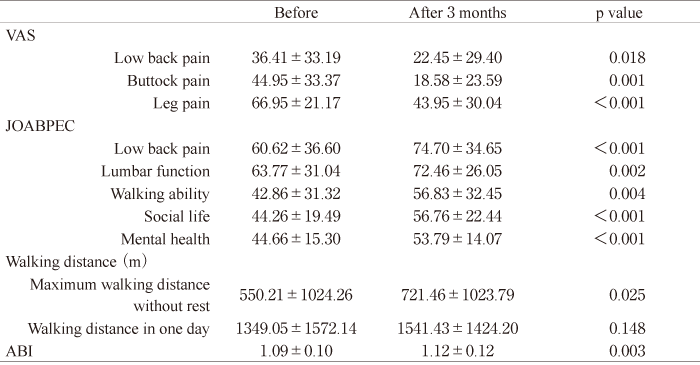
Changes in pain scores and walking distance VAS scores for low back pain, buttock pain, and leg pain were 36.41±33.19, 44.95±33.37, and 66.95± 21.17, respectively, before administration of limaprost, and 22.45±29.40, 18.58±23.59, and 43.95±30.04, respectively, after administration of limaprost for 3 months. Each score showed a significant improvement after treatment with limaprost(P<0.01)(Table 2). JOABPEQ scores for the 49 patients in 5 categories(low back pain, lumbar function, walking ability, social life function, and mental health) are also shown in Table 2. All categories significantly improved after 3 months of limaprost treatment(low back pain: P<0.001, lumbar function; P=0.002, walking ability: P=0.004, social life function: P<0.001, and mental health: P<0.001). There was also a significant improvement in maximum walking distance without rest from before to after treatment(550.21±1024.26 vs. 721.46±1023.79 m, P=0.025), but there was no significant improvement in walking distance in one day after treatment(P=0.148)(Table 2)
Table 2
Pain scores, walking distance and ABI before and after 3 months of treatment with limaprost
In the current study, limaprost improved low back pain, leg pain, and walking difficulty that originated from lumbar spinal stenosis, and also increased ABI. These findings indicate that limaprost is useful to treat symptoms of lumbar spinal stenosis and to increase blood flow in the anterior and posterior tibial arteries. PGE1 is a vasodilator that increases blood flow and inhibits platelet aggregation[5]. Intravenous PGE1 is primarily used for chronic peripheral arterial occlusive diseases in the United States and Europe[5]. PGE1 also increases blood flow at the surface of spinal nerve roots, and thus PGE1 analogs have been developed to treat symptoms of lumbar spinal stenosis[5,17]. These drugs include limaprost, an oral PGE1 analog that was developed in Japan. The efficacy of oral limaprost was evaluated in adult Japanese patients in 3 randomized, double-blind, 6-week trials[17]. Limaprost at a dose of 15 μg/day was superior to a dose of 3 μg/day for overall efficacy and improvement from baseline in a phase III trial in 146 patients with lumbar spinal stenosis. The efficacy of limaprost at 15 μg/day did not differ significantly from that at 30 μg/day, so the optimal dosage of limaprost for this indication was concluded to be 15 μg/day[17]. In a comparative randomized control trial, Matsudaira et al. found that limaprost was superior to etodolac, a NSAID, for health-related quality of life in patients with symptomatic lumbar spinal stenosis with cauda equina symptoms[18]. Consistent with these findings, in the current study limaprost improved pain and walking difficulty originating from lumbar spinal stenosis.
In the current study, we administered limaprost for 3month. Because, we considered that it needed longer period for improvement of neurologic symptom form lumbar spinal stenosis compared to improvement of blood flow in vessels. Indeed, Matsudaira et al. found limaprost was administered for 8 weeks and improvement of symptom in the patient with lumbar spinal stenosis[18]. We concluded this observational period was valid for these reasons.
Limaprost also increased ABI in our patients. For PAD, the recommended pharmacotherapy includes cilostazol, and PGE1 has been reported to improve the functional capacity of patients with claudication[10]. These drugs also improve maximal walking distance by 40% to 60%[19,20]. These therapies generally increase flow in capillary vessels and do not affect the main arterial tract. For this reason, most reports have shown that these drugs do not improve ABI[4,12]. In contrast, Mohler et al. found that treatment of PAD with cilostazol significantly improved exercise performance and increased ABI, although with the conclusion that the mechanism underlying the improved ABI was unclear[21]. Compression of the spinal nerve roots has been shown to induce degeneration of nerves and reduce blood flow in nerve roots in animal models and humans [1,2]. Spinal ventral nerve roots contain unmyelinated nerve fibers associated with sensory and autonomic nerves, and myelinated nerve fibers associated with motor fibers[13-15]; and somatosensory, somatomotor, and autonomic nerve fibers run on the surface of spinal nerves and the dura mater[15]. Animal and human cadaver studies have shown that lumbar and sacral spinal ventral nerve roots contain many unmyelinated nerve fibers and sympathetic and parasympathetic nerves that innervate blood vessels in the lower leg[13,14], with no significant difference in the proportions of unmyelinated nerve fibers among sympathetic(T11-L2), parasympathetic(S2) and other(C4-T10 and L3-S1) segments[14]. Sympathetic and parasympathetic nerve fibers may be damaged and compressed in patients with lumbar canal stenosis, and increased blood flow to the spinal nerve roots caused by limaprost may result in recovery of function of the autonomic nervous system. Thus, this may explain the increase in ABI after treatment with limaprost in the current study.
There were several limitations in the study. First, we did not use a control group taking NSAIDs only. Second, a normal ABI falls in the range of 0.91-1.30, while a low ABI at rest(<0.90) indicates a high risk of PAD[22]. In the current study, most patients had ABI in the normal range and the clinical significance of an increase of ABI in this population is questionable. Therefore, a further study is needed to test our hypothesis more rigorously. Within these limitations, we conclude that limaprost improved low back and leg pain originating from lumbar spinal stenosis, and increased ABI scores. These findings indicate that limaprost is useful to treat symptoms caused by lumbar spinal stenosis and blood flow in the main artery in the lower leg.
The author declares no conflicts of interest with respect to the authorship and publication of this article.
The author received no financial support for the research or authorship of this article.
Address correspondence to Dr. Seiji Ohtori.
Department of Orthopedic Surgery, Graduate School of Medicine, Chiba University, 1-8-1 Inohana, Chuo-ku, Chiba 260-8670, Japan.
Phone: +81-43-226-2117. Fax: +81-43-226-2116.
E-mail: sohtori@faculty.chiba-u.jp
