


Chiba Medical J. 89E:9~14,2013
doi:10.20776/S03035476-89E-2-P9
[Original Paper]
Kazuya Yamaguchi, Kanae Mitsuhashi, Yuki Nakagawa and
Manabu Hayashi
Comprehensive Medical Checkup Center, Chiba Foundation for Health Promotion and Disease Prevention, Chiba 261-0002.
(Received January 4, 2013, Accepted February 8, 2013)
Background: Comfort during colonoscopy is a universal concern, but when the patient is drugged and unable to change position, this can prejudice the accuracy of colonoscopy.
Aim: To assess whether position changes during the colonoscope withdrawal phase decrease the sensation of abdominal fullness.
Methods: This study involved 150 patients, none of whom received intravenous sedation. After colonoscopy to the cecum in the supine position, the examinations were completed with the following dynamic position changes( cecum to transverse colon, supine; splenic flexure and descending colon, right lateral; sigmoid colon, supine; and rectum, left lateral) or minimal position changes( cecum to sigmoid colon, supine; and rectum, left lateral). Patients graded the sensation of abdominal fullness as follows: Grade 1: no abdominal fullness; Grade 2: mild fullness; Grade 3: moderate fullness; Grade 4: marked fullness; and Grade 5: painful fullness.
Results: The following scores were reported with dynamic position changes: Grade 1, 86.7%; Grade 2, 12.0%; Grade 3, 1.3% and with minimal position changes Grade 1, 53.3%; Grade 2, 44.0%; Grade 3, 2.7%. Dynamic position changes significantly decreased patient-assessed abdominal fullness grades compared with minimal position changes( P<0.05) according to the Mann-Whitney U test.
Conclusions: Dynamic position changes during colonoscope withdrawal decrease the sensation of abdominal fullness.
Colonic diseases, Colonoscopy, Endoscopy, Insufflation, Patient positioning
In recent years, various approaches to improve colonoscopic quality have been advocated. However, the basic colonoscopic technique can benefit from reevaluation [1]. Withdrawal times of ≥ 6 minutes, monitoring and feedback programs to improve the detection rates for small adenomas and large( ≥ 10 mm) adenomas, and the use of position changes during colonoscope withdrawal can reduce the amount of colonic surface area missed [2-4].
Routine position changes during the withdrawal phase of colonoscopy are not widely practiced, the entire procedure often being performed in the left lateral position for convenient access. In this position, gravity causes the capacious and fragile right colon to distend first. According to Laplace's law, for any given intraluminal pressure, greater right colonic diameters result in greater wall tension and increased potential for damage. Unless carbon dioxide insufflation is used, overdistension more commonly simply results in postprocedure discomfort [5].
Because performing position changes is difficult in heavily sedated or anesthetized patients, any advantages of position change are so far undetermined [4]. The use of position changes during the withdrawal phase of colonoscopy was initially based on the experience of radiologists administering barium enemas: they discovered that moving the patient facilitated adequate distension of the colon and movement of excess fluid away from the colonic area of interest. Anatomically, this relies on having the region of interest at the highest point, causing gas to rise and fill the area and fluid to fall away to dependent areas. East et al. investigated the role of dynamic position changes during colonoscope withdrawal in luminal distension. Their study demonstrated that changing the patient’s position to supine for examination of the transverse colon and to the right lateral position for examination of the splenic flexure and descending colon during colonoscope withdrawal significantly improved colonic luminal distension in these areas and led to a significant decrease in the proportion of patients with diagnostically unacceptable distension [4]. Such improvement in luminal distension may improve adenoma and early carcinoma detection rates [6].
Our aim was to investigate whether position changes during the colonoscope withdrawal phase decrease abdominal fullness.
Subjects
Between December 2009 and July 2010, 150 patients aged 36 to 80 years who had intact colons and had presented for routine colonoscopy at our center were invited to take part in the study (Table 1). Each of the patients gave written informed consent before entry into the study and the study was approved by the Ethics Committee of our center. We received no funding or material support for this trial.
Table 1
Patient characteristics (n=150)
Colonoscopy
Patients received intramuscular antispasmodic medication unless contraindicated and did not receive intravenous sedation unless requested (Table 1). Colonoscopes were CF 260 series variable-stiffness instruments(CF-Q260AI, CF-H260AI, CF-H260AZI) (Olympus Medical Systems Corporation, Tokyo, Japan). All colonoscopies were performed by three experienced operators(more than 3000 colonoscopies performed). Patients were randomized prior to the procedures to examination with either dynamic or minimal position changes. Sealed envelopes were used to randomly allocate patients to these two groups. As much fluid and air as possible was suctioned out during intubation. Because they had been excessively insufflated with air and carbon dioxide, patients were excluded if the examination was not completed within 40 minutes(classified as technically difficult).
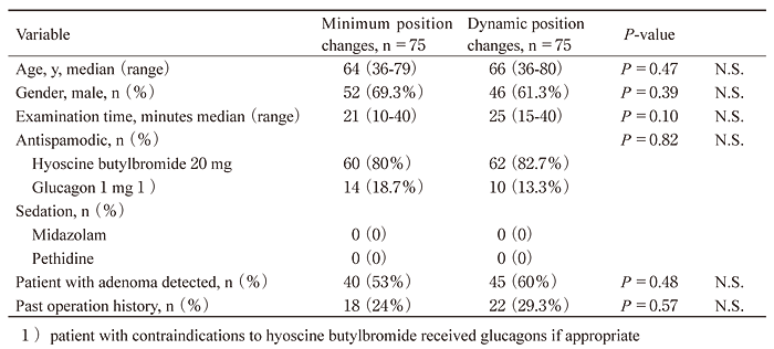
After cecal intubation, colons were examined in five segments:(1) cecum, ascending colon;(2) transverse colon;(3) descending colon;(4) sigmoid colon; and (5) rectum. Examination in the supine position was followed by either the following dynamic position changes(cecum to transverse colon, supine; splenic flexure and descending colon, right lateral; sigmoid colon, supine; and rectum, left lateral) (Figure 1) or minimal position changes( cecum to sigmoid colon, supine; and rectum, left lateral)) (Figure 2). After examination of each segment, as much air as possible was suctioned out. The position of the colonoscope in each segment was determined clinically by the endoscopists as previously described[4].
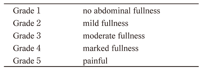
Carbon dioxide was continuously insufflated with an endoscopic CO2 regulator( UCR, Olympus Medical Systems Corporation) to improve the range of vision while each area was examined. Polyps over 3 mm were biopsied. An experienced GI pathologist examined all excised polyps. After examination, patients were questioned about abdominal fullness and asked to score it as one of the following grades: Grade 1: no abdominal fullness; Grade 2: mild fullness; Grade 3: moderate fullness; Grade 4: marked fullness; Grade 5: painful (Table 2).
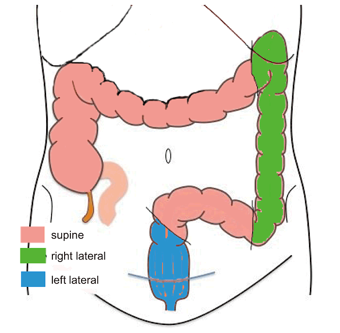
Fig.1
Position changes during the colonoscope withdrawal phase with dynamic position change sequence.
Fig.2
Position changes during the colonoscope withdrawal phase with minimal position change sequence.

Fig.3
Participant flow through the trial.
Table 2
Grades of the sensation of abdominal fullness
Statistical analysis
Age, examination time and patients with adenoma detection were compared using the F test and the Student’s t-test. Other variables were compared using the F and Mann-Whitney U tests. The differences in abdominal fullness scores between minimal position changes and dynamic position changes were compared using the F, Mann-Whitney U and Welch’s t-tests. A P value <0.05 was considered statistically significant. P values between 0.05 and 0.10 were considered a statistical trend.
All statistical calculations were performed using Statcel(OMS, Tokorozawa, Saitama, Japan).
Based on our expectation that few people would nominate Grades 3, 4, or 5, it was determined that 150 patients would be needed to complete the study. If large numbers of patients nominated Grade 5(painful), we planned to end the trial.
Patients’ Characteristics
The characteristics of the patients receiving carbon dioxide insufflation are presented in Table 1 (Table 1). According to the Mann-Whitney U test, there was a small, nonsignificant difference in adenoma detection rates(P= 0.48). We found more adenomas with dynamic position changes than with minimal position changes.
Examinations with carbon dioxide insufflation
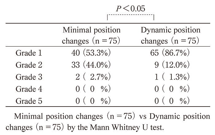
For dynamic position changes, patients reported the following scores: Grade 1, 86.7%; Grade 2, 12.0%; Grade 3, 1.3%; Grade 4, 0%; and Grade 5, 0%. For minimal position changes, they reported the following scores: Grade 1, 53.3%; Grade 2, 44.0%; Grade 3, 2.7%; Grade 4, 0%; and Grade 5, 0%. Thus, 86.7% of patients with dynamic position changes and 53.3% of those with minimal position changes nominated Grade 1(no abdominal fullness). No patient in either group reported Grade 5(painful). Dynamic position changes significantly decreased abdominal fullness grades compared with minimal position changes(P<0.05) according to the Mann-Whitney U test (Table 3).
There were no adverse events related to implementation of position changes during the study.
Table 3
Score of abdominal fullness
Methods reported to reduce discomfort during colonoscopy include the use of pediatric colonoscopes, variable stiffness colonoscopes, or gastroscopes; inhalation of nitrous oxide; insufflation with carbon dioxide; hypnosis; music; audio distraction; or simply allowing the patients to participate in administration of their sedation [7]. A combination of the water method with on-demand sedation minimizes patient recovery-time [8]. However, no previous reports have discussed the relationship between position changes during colonoscopy and post-examination abdominal fullness. To perform position changes easily and quickly, we do not routinely use sedation. Attention to straightening the colonoscope, suctioning fluid and extra gas, and using position changes during both insertion and withdrawal phases allows for minimum sedation and a comfortable examination.
Endoscopists can use gas insufflation and antispasmodics in addition to position changes to improve luminal distension. Nearly all patients in our study received the antispasmodic hyoscine butylbromide or glucagon to pharmacologically maximize distension; we use these routinely based on expert experience. Because nearly all patients received antispasmodics, the benefit of position changes is additive to that of antispasmodics. We used carbon dioxide for insufflation; this has repeatedly been shown to cause less discomfort than air, be safe, and likely contribute to improved acceptability of colonoscopy. In previous studies, 15% of patients felt pain when carbon dioxide was used for insufflation [5].In our study, with both dynamic and minimal position changes, no patient reported feeling pain( Grade 5)
We did not measure the amount of carbon dioxide introduced during examination with either dynamic position changes or minimal position changes, because the amount of gas or fluid suctioned and the very rapid absorption of carbon dioxide across the bowel wall (100 times faster than air) are significant influencing factors [9,10]. If insufflation is continued for long periods because of sustained poor luminal distension, high pressures can build up in the colon, requiring active deflation by reintubation and suction after completion of the examination. By limiting the need for gas insufflation to achieve an adequate luminal view, position change during withdrawal may also improve patient comfort and safety.
Despite randomization, both patients and operators knew to which group they were assigned( dynamic or minimal position changes). Therefore, our trial was not completely blinded.
Position change is likely to be a cost-neutral intervention. Additionally, position change has no adverse effects and can easily be used in almost all patients who are no more than lightly sedated. It does not require extra equipment or training and takes about 30 seconds per change. In our center, dynamic and minimal position changes are routinely used, the operator making the choice between these. Dynamic position changes require an additional 5 minutes per case for the four changes( left lateral to supine, supine to right, right to supine, and supine to left). This additional time does not impact unit efficiency and may even save time overall; less time may be spent insufflating and suctioning fluid pools and discharge may be faster because there is less gaseous distension after the procedure. The improved luminal distension may improve adenoma and early carcinoma detection rates [6,11-13]. Integration of position changes into routine clinical colonoscopic practice has the potential to improve efficacy of national bowel cancer screening programs without requiring additional resources.
In this study, we suggest that dynamic position changes during colonoscope withdrawal, supine to right lateral for the splenic flexure and the descending colon, improves distension of the lumen compared with minimal position changes and supine position for examination of these areas. Additionally, the reduced air and carbon dioxide insufflation required for viewing leads to decreased abdominal fullness. This study confirms anecdotal and expert opinion on the role of position changes during colonoscopy withdrawal. Dynamic position changes could easily become a basic colonoscopic technique.
We thank the staff of the Gastroenterology Division, Comprehensive Medical Checkup Center, Chiba Foundation for Health Promotion and Disease Prevention.
We received no funding or material support for this trial.
None
Address correspondence to Dr. Kazuya Yamaguchi.
Gastroenterology Division, Comprehensive Medical Checkup Center, Chiba Foundation for Health Promotion and Disease Prevention, Mihama-ku, Chiba 261-0002, Japan
Tel.+81-43-246-0350. Fax. +81-43-246-8640.
E-mail: ka-yamaguchi@kenko-chiba.or.jp
