| School
of Medicine, Chiba University Chiba University Hospital |
|
|
|
|
|
| Chiba Medical Journal | |
| Journal Index | |
| 〔Original Paper〕 | ||||
| Correction of Experimentally Induced High Myopia Inhibits Recovery by Spectacle Lenses in Chicks |
||||
|
|
||||
| Akira Ito1), Hidehito Kawabata2) and Emiko Adachi-Usami3) | ||||
| (Received December 4, 2006, Accepted December 12, 2006) | ||||
|
|
||||
SUMMARY
|
||||
Key Words
|
||||
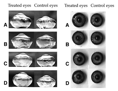
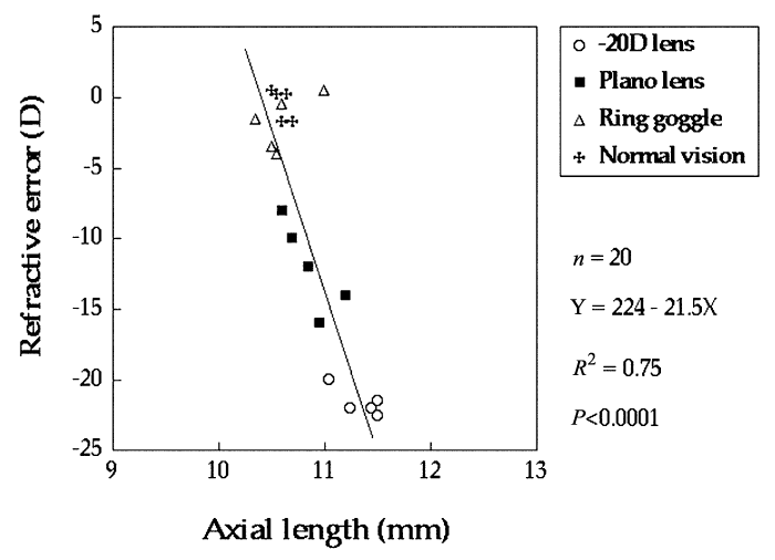
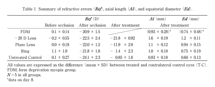
| I. Introduction The retina is not only a sensory organ of vision but also an effector organ that controls eye growth by feedback mechanisms in the retina[1]. Emmetropization, the nulling of refractive errors or the process of matching axial length to the focal plane, is an interesting and practical process observed in many species[2-5]including humans[6]. Form-deprivation induces axial myopia but the myopia can be stopped and reversed if the form-deprivation is eliminated[7-9]. When the retinal image is defocused by optical lenses the eye grows to attain an image focused on the retina and the growth is altered when the lenses are removed in chicks[10-12], tree shrews[13], and monkeys[14]. There are two major mechanisms involved in emmetropization[1,15]. The first is a vision-dependent mechanism with visual feedback to guide the eye to emmetropia by controlling axial growth. Wildsoet et al showed that the recovery from deprivation myopia is prevented by wearing appropriate lenses in chick[16], and McBrien et al obtained similar results in the tree shrew[17]. The other mechanism is a non-vision-dependent, shape-sensitive mechanism programmed to return the eye to its normal shape. In order to examine these mechanisms, we induced about -20 D of high myopia by form deprivation in chicks, and determined whether recovery from the experimentally-induced high myopia can be prevented by correcting the myopia by appropriate spectacle lenses in young chicks. II. Materials and Methods One-day-old male White Leghorn chicks (Gallus domesticus) were obtained from a local hatchery (Togane, Japan) and were raised in the laboratory under fluorescent lighting set to a 12/12 hour light/dark cycle. The illumination was about 300 lux at the level of the cages. All chicks had access to water and food ad libitum, and the cages were cleaned twice a day. We also weighed all animals and found that all animals grew at approximately the same rate. The treatment and care of the chicks were in accordance with the ARVO Statement on the Use of Animals in Ophthalmic and Vision Research. To induce form-deprivation myopia, we constructed translucent occluders from laboratory translucent polypropylene test tubes (16 mm diameter). Forty chicks were monocularly deprived of sharp vision by gluing these translucent occluders to the feathers around the treated eyes for a week (from day 1 to day 8). Our preliminary experiments showed that a week of form-deprivation induced approximately -20 D of high myopia in the chicks. We selected 25 chicks that became myopic by about -20 D from forty chicks. The contralateral non-deprived eyes served as controls. We constructed ring goggles, giving a field of view of approximately 90-100 deg, from the occluders, and lenses were attached to the ring goggles. The lenses (Hoya Healthcare Corporation, Tokyo, Japan) were made from PMMA material (7.80 D base curve: 10.0 mm over all diameter) which were either -20 D or zero-powered, and they were mounted on the ring goggles. After determining the refractive error on day 8 (after one-week form-deprivation), five chicks, Group FDM, were set aside for measurements of the eye dimensions. The other twenty animals were randomly divided into four treatment groups (n=5 in all groups). Group -20 D wore -20 D lenses and Group PL wore plano lenses attached to the ring goggles in front of the treated eyes. The outer surface of lens was cleaned twice a day. Group RG wore empty ring goggles in front of the treated eyes to determine the effect of the ring goggles. Group NV restored normal vision following one-week form-deprivation. Cycloplegia was obtained with repeated application of topical 0.2% pancuronium bromide, muscle relaxant[18-20]; every 15 minutes in a dark room and refractive error was measured 1 hour later. Refractive error of the chick eyes was measured along the optic axis in the horizontal meridian by retinoscopy. Measurements were made three times during the course of the experiments: before occlusion (day 1), after form-deprivation (day 8), and after treatment (day 15, except for Group FDM). All refractions were performed by one of the authors (H. K.), who is experienced in doing retinoscopy on small eyes. After retinoscopy on day 15 (or on day 8 for Group FDM), animals were killed with an overdose of urethane and the eyes were enucleated for the measurements of the intact globe. The eyes were cleaned of all extraocular tissues, and axial length and equatorial diameter were measured with a slide caliper (Fig. 1). The interocular differences between the treated and contralateral control eyes (T-C differences) in the refraction and eye dimensions were calculated and used for the statistical analysis. The differences between Group FDM and four treatment groups were analyzed using unpaired t-test (significant level was 0.05). The differences among four treatment groups were determined by one-way analysis of variance with Bonferroni/Dunn’s correction of P value for multiple comparison (statistical significant level was 0.0083). We also evaluated the coefficient of correlation between the refractive error and the eye dimensions in the treated eyes on day 15 using simple regression analysis (significant level was 0.05). III. Results In Groups PL, RG and NV, the interocular difference (T-C difference) in the refraction on day 15 was significantly reduced than that on day 8, respectively. Thus, not correcting the induced myopia by lenses led to a recovery from the myopia in Groups RG and NV and a partial recovery in Group PL. (Table 1) However in Group -20 D, the T-C difference in the refraction on day 15 did not change significantly from that on day 8 (P=0.41). This showed that the correction of the induced high myopia by an appropriate spectacle lens inhibited the recovery from the myopia. The T-C difference in the refraction in Group -20 D was significantly greater than that in Group PL, Groups RG and NV (P<0.001) (Table 1). On day 8, the T-C differences of the axial and equatorial lengths showed that there had been excessive growth in both dimensions in the form-deprived eyes (Group FDM). On day 15, the T-C difference in the axial length of Group -20 D had increased further compared of Group FDM (P<0.01), and the differences were significantly larger than that of the other three groups (P<0.001). The T-C differences between Groups PL, RG and NV were not significant (Table 1). On day 15, the T-C differences in the equatorial diameter of Group -20 D had increased compared of Group FDM (P<0.01), and the T-C difference of Group -20 D was significantly greater than that of the other three groups (P<0.001). The T-C differences between Groups PL, RG and NV were not significant (Table 1). On day 15, the coefficient of correlation between the refractive error and the axial length of the treated eyes was highly significant with R2=0.75 (n=20, Fig. 2). | ||||
|
||||
|
||||
| IV. Discussion These results demonstrated that appropriate correction of the induced high myopia inhibited recovery of the induced myopia and the myopia was maintained (Group -20 D). On the other hand, not correcting the myopia led to a recovery from the myopia (Group RG and NV) and a partial recovery by plano lens (Group PL). This result is in agreement with earlier reports on chicks [16] and tree shrews [17] . Thus, these findings support an active vision-dependent emmetropization factor even in -20 D of high myopia. It should be noted that we did not find a shape-sensitive mechanism acting to change the shape of the eyes to normal. Even though the T-C differences of Group -20 D were increased in relative axial length compared to Group FDM, the T-C differences in refractive error for Group -20 D did not change suggesting that there may have been some choroidal compensation [18-20] to stabilize their refractive error. The induced myopia of the Group PL did not completely recover as myopia was still found on day 15. This myopia could be due to a mild deprivation caused by a small quantity of food attached to the lens surface [21] which could have blurred retinal image slightly and thus prevented the complete recovery. There was no significant difference in the refractive error or the eye dimensions between Group RG and NV. This indicates that the ring goggle wear was not critical, and the diameter of the open ring goggle and the lens-mounted goggle was large enough for chick’s visual field as far as in this experiment. It is interesting that Group RG and NV showed recovery in the refractive error but the absolute interocular differences were still existed. A substantial choroidal compensation [18-20] during recovery in only a week may be the explanation for this discrepancy. In conclusion, our observations showed that appropriate refractive correction of the experimentally-induced high myopia in neonatal chicks prevented the recovery from the myopia and thus supports the concept of visually dependent eye growth regulation. V. Acknowledgements The authors thank Professor Duco Hamasaki for his helpful comments and editorial support in manuscript preparation. This study was supported in part by a Grant in-aid for scientific research (No. 10357015) from the Ministry of Education, Science, Sports and Culture, Japan. | ||||
|
|
||||
要旨
|
||||
References
|
||||
Others
|
||||
|