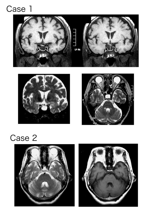
Fig. 2
MR Images of the brain of Cases 1 and 2. Note the empty enlarged sella turcica and tilted chiasma in Case 1. The upper figures are T1 and the lower figures are T2 images in Case 1. The left figure is T2 and the right figure is T1 image in Case 2.