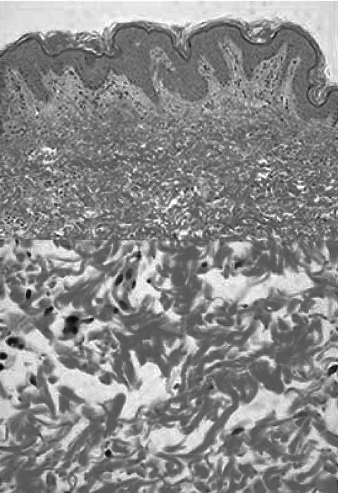
Fig. 2
Histological skin sections of case 1. Under low magnification, dermal collagen fibers appeared rough and aberrant. Under high magnification, the thickness of collagen fibers was uneven and ruptured fibers were seen.
Fig. 2
Histological skin sections of case 1. Under low magnification, dermal collagen fibers appeared rough and aberrant. Under high magnification, the thickness of collagen fibers was uneven and ruptured fibers were seen.