


Volume 84, Number 3
doi:10.20776/S03035476-84-3-P121
[Original Paper]
Seiji Ohtori, Hiroaki Sameda, Yasuaki Murata, Eiji Hanaoka, Shinichiro Nakamura
Yuzuru Takahashi, Masatsune Yamagata and Kazuhisa Takahashi
(Received December 26, 2007, Accepted January 23, 2008)
To clarify the afferent pathways from intervertebral discs to dorsal root ganglia (DRGs) in humans we evaluated the effect of an upper and lower spinal nerve block on discogenic low back pain. Patients suffering from discogenic low back pain originating from the L4/5 or L5/S1 intervertebral disc received a spinal nerve root block (L2 nerve block group: n=34, L4 or L5 nerve block group: n=34). Lidocaine (1.5 ml of 1% solution) was administrated to L2, L4, or L5 spinal nerves. In both groups, spinal nerve blocks were significantly effective in alleviating discogenic low back pain (P<0.05). Fifteen minutes after the block, the average visual analogue pain scale score decreased from 8.0 to 4.3 (L2 root block group) and from 7.8 to 3.4 (L4 or L5 root block group). The average effective period was significantly longer in the L2 root block group (13 days) than in the L4 or L5 root block group (8 days) (P<0.05). The upper and lower spinal nerves appear to include sensory afferent nerves from the L4/5 or L5/S1 intervertebral disc. There were differences in the intensity and the period of effective relief between the upper and lower nerve block.
Dorsal root ganglion, Intervertebral disc, Pain, Spinal nerve
In the human lumbar intervertebral disc, many studies have described the existence of sensory nerve endings in the annulus fibrosus [1]. It has been believed that such nerve endings originate from the sinuvertebral nerves branching from the ventral ramus of the spinal nerve and the ramus communicans of the corresponding level in human [1]. Recent studies have revealed that the dorsal portion of the L5/6 intervertebral disc is multi-segmentally innervated by dorsal root ganglia (DRGs) from T13 to L6 levels in rats [2,3]. Some rat sensory nerve fibers from the L5/6 intervertebral disc pass to upper DRGs via paravertebral sympathetic trunks [2,3]. However, in humans the sensory afferent pathway from the lower intervertebral disc has not been clarified. Nakamura et al. performed an L2 spinal nerve block based on animal sensory innervation and reported that the block was effective to patients suffering from discogenic low back pain [4]. However, they did not compare this with nerve blocks at different levels. The aim of this study was to evaluate and compare the effect of both upper and lower spinal nerve blocks on L4/5 or L5/S1 discogenic pain in humans, and to suggest the sensory pathway from the L4/5 intervertebral disc to the DRGs.
The protocols for human procedures in these experiments received approval from the ethics committees of our institutions.
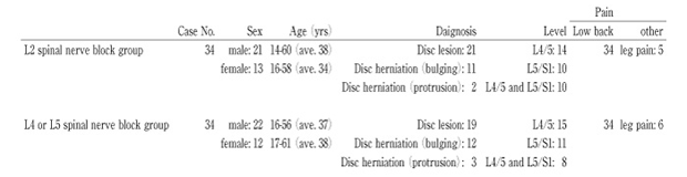
We studied 68 patients (43 male and 25 female) whose average age was 36±4 years (Mean±S.E.M.) (range 14〜61 years). Details are shown in Table 1. They all suffered from low back pain. In eleven patients this also involved leg pain. Discogenic pain was diagnosed using physiological examination, pain location, radiography, and magnetic resonance imaging (MRI). We examined patients whose low back pain was exacerbated by forward bending of the trunk as described by Nakamura et al. [4]. We also examined patients whose only indication was L4/5, L5/S1, or both intervertebral discs degeneration on MRI imaging. Patients who had severe spinal spodylolysis, or disc degeneration with three or multi-level lesions were excluded. Patients who had received spinal surgery were also excluded.
Administration was as described in a previous report [4]. On the predominantly painful side, a 22-guage spinal-nerve-block needle was advanced obliquely to the corresponding spinal nerve under fluoroscopic control (L2 spinal nerve block group, n=34; L4 or L5 spinal nerve block group, n=34). In L4 or L5 spinal nerve block group, L4 or L5 spinal nerve block was applied to only L4/5 disc degeneration, and L5 spinal nerve block was applied to only L5/S1 disc degeneration. If the patients had two disc degeneration (L4/5 and L5/S1), L4 spinal nerve block was applied to the patients. Then 0.5 ml of the contrast medium Iotorolan (Schering AG, Berlin, Germany) was injected to confirm the position of the spinal nerve. Unilateral lidocaine administration (1.5 ml of 1% solution) was then performed. The intensity of low back pain was evaluated before the block using a visual analogue scale (VAS; score, 0〜10: a score of 10 being the worst pain). At 15 minutes, 7 days, 14 days, and 21days after spinal nerve block, the VAS scores were also examined. We defined periods during which patients indicated less than 60% of their VAS score before nerve block as effective periods. The differences between the groups were compared by one-way analysis of variance (ANOVA) for repeated measurements. Differences were considered to be statistically significant at P<0.05.
Table 1
Details of 68 patients with discogenic pain
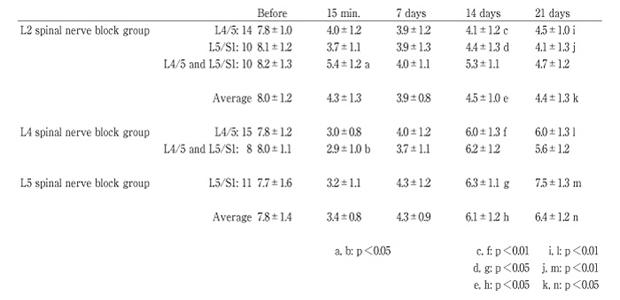
VAS scores before blocks ranged from 7.2 to 10 (8.0±1.2; Mean±S.E.M) in the L2 spinal nerve block group, and from 6.8 to 10 (7.8±1.4; Mean±S.E.M) in the L4 or L5 spinal nerve block group. In both groups, spinal nerve blocks were significantly effective in attenuating discogenic low back pain at 15 minutes, and 7, 14, and 21 days after block (P<0.05) (Table. 2). One patient in the L2 nerve block group and one patient in the L4 or L5 spinal nerve block group were not alleviated of low back pain after the nerve block. Average VAS scores were 4.3±1.3 (15 minutes), 3.9±0.8 (7 days), 4.0±1.0 (14 days), and 4.4±1.3 (21 days) in the L2 spinal nerve block group and 3.4±0.8 (15 minutes), 4.3±0.9 (7 days), 6.1±1.2 (14 days), and 6.4±1.2 (21 days) in the L4 or L5 spinal nerve block group. There was no significant difference between the groups at 15 minutes after the blocks (P>0.1). The average VAS score was lower at 14 and 21 days in the L2 spinal nerve block group than in the L4 or L5 spinal nerve block group (P<0.05). VAS scores in the L2 spinal nerve block group for L4/5, L5/S1, or L4/5 and L5/S1 disc degeneration were not significantly different from each levels (P>0.05) (Table. 2). VAS scores in L4 spinal nerve block for L4/5, L5/S1, or L4/5 and L5/S1 disc degeneration were not significantly different from those in L5 spinal nerve block for each level (P>0.05) (Table. 2). The average effective period was significantly longer in the L2 root block group (13 days) than in the L4 or L5 root block group (8 days) (P<0.05).
Table 2
Details of VAS after block
In the current study, discogenic pain was diagnosed using pain location, physiological examination and MRI. It is generally difficult to prove the origin of low back pain. However, we defined low back pain as originating from intervertebral discs using the forward bending trunk test [4]. MRI findings that excluded the other diagnosis of low back pain substantiated our method. The defined pain was alleviated by spinal nerve block at several different levels.
Intervertebral discs have long been thought to be the main source of the common form of low back pain [5]. Many findings regarding discogenic pain in humans and animals have been reported.
Low back pain has been produced by intra-operative stimulation of the outer annulus or the posterior longitudinal ligament under local anesthesia in human [6]. Discography induces low back pain in human [7]. Many studies have demonstrated nerve endings in the annulus fibrosus in the intervertebral disc [1]. Recently, many investigators have identified nerve fibers, immunoreactive for the putative nociceptive markers substance P or calcitonin gene-related peptide (CGRP), in intervertebral discs and DRG neurons innervating intervertebral discs [8,9].
Low back pain is usually increased in sustained sitting positions, especially involving forward bending of the trunk, both of which are known to increase intradiscal pressure [10]. We assume that the forward bending trunk test, which was used in this study, increases the intradiscal pressure and distends the outer layer of the annulus. As a consequence some sensory nerves in the annulus are stimulated.
In rats, sensory nerve fibers from the L5/6 intervertebral disc were thought to pass through the sinuvertebral nerves on the posterior longitudinal ligament and reach the DRGs from L3 to L6. In the non-segmental innervation, sensory nerve fibers were thought to enter the paravertebral sympathetic trunks through the L5 ramus communicans and reach the DRGs from T13 to L2 directly through each ramus communicans [2,3,11,12]. In humans, the sensory afferent pathways remain unclear. However, some data have suggested the same pattern of sensory nerve innervation. In the case of lumbar intervertebral disc degeneration, blockade of the spinal nerves at the same level has been effective for some patients with discogenic disorders, but for other patients blockade of L2 spinal nerves or paravertebral sympathetic trunks is effective [4,13,14]. In the current study, we did not perform spinal nerve blocks at all levels. However, at least sensory nerves originating from L2, L4, and L5 appear to innervate the human L4/5 or L5/S1 intervertebral disc. In humans, some DRGs at different levels seem to innervate the L4/5 or L5/S1 intervertebral disc. We believe that the sensory pathway from the upper DRG to the disc is via paravertebral sympathetic trunks.
The difference in the effective period of nerve block between the L2, L4, and L5 nerves may be rationalized by the following reports. The number of L2 DRGs innervating the rat L5/6 intervertebral disc is more than that of L4 or L5 DRG [2,3]. In addition to the intervertebral disc, lumbar muscles and lumbar facet joints also received sensory nerve fibers from L1 or L2 DRGs via paravertebral sympathetic trunks [15-17]. Some sympathetic neurons innervating the lumbar facet joint connect with CGRP-immunoreactive sensory fibers through synaptic contact in paravertebral sympathetic trunks [16]. This would allow the transmission of sensory information to sympathetic postganglionic neurons innervating lower lumbar facet joints. Sympathetic postganglionic fibers in the paravertebral sympathetic tract can exert an influence on the activities of the dorsal horn sensory neurons receiving nociceptive stimuli from lumbar paraspinal tissues [15] and some sympathetic postganglionic fibers regulate the metabolism of vertebral bone and connective tissues [16]. They also may be related to sensory activity in lower lumbar facet joints. If the nervous connection between sympathetic postganglionic neuron innervation in the human intervertebral disc and CGRP-immunoreactive sensory fibers is the same, this would be consistent with the difference in the effective period after L2, L4, and L5 spinal nerve block.
1) We defined low back pain as originating from intervertebral discs using the forward bending trunk test and MRI findings. Discography was necessary for diagnosis of discogenic low back pain.
2) Unilateral 1.5 ml of lidocaine administration was performed for L4 or L5 nerve root block group, and the nerve block was effective. We can not deny leakage of lidocaine into intervertebral disc in L4 or L5 nerve root block group.
We need further study to clarify these points.
【目的】
ヒト椎間板を支配する後根神経節からの感覚神経支配を調べるために,椎間板性腰痛に対して,上位の神経根ブロックと下位の神経根ブロックを比較検討したので報告する。
【方法】68人のMRIにてL4/5またはL5/S1に椎間板変性を伴う腰痛患者に対し,ランダムに神経根ブロックを行った。内訳はL2神経根ブロックが34人,L4またはL5神経根ブロックが34人であった。それぞれ,1.5mlのリドカインを使用し,ブロック前後の疼痛と効果期間に関してvisual analogue pain scale (VAS) を用いて評価した。
【結果】
ブロック後,両群共に椎間板性腰痛を有意に軽減させた (P<0.05) 。ブロック前に比較し,ブロック15分後のVAS値はL2神経根ブロック群で8.0から4.3に,L4またはL5神経根ブロック群で7.8から3.4と改善していた。ブロック前後のVAS値に関し,両群に差はなかった (P>0.05) 。しかしながら,ブロックの効果有効期間はL2神経根ブロック群で平均13日,L4またはL5神経根ブロック群で平均8日であり,有意にL2神経根ブロック群に効果が長かった (P<0.05) 。
【考察と結論】
上位後根神経節由来 (L2神経根) ,下位後根神経節由来 (L4またはL5神経根) の感覚神経線維は共に,L4/5またはL5/S1椎間板を支配していると考えられた。ブロックの効果に関して,L2神経根ブロック群とL4またはL5神経根ブロック群で効果期間に有意差があった。その機序は不明であるが,今後更なる検討が必要と考えられた。
Department of Orthopedic Surgery, Graduate School of Medicine, Chiba University, Chiba 260-8670.
大鳥精司,鮫田寛明,村田泰章,花岡英二,中村伸一郎,高橋 弦,山縣正庸,高橋和久: ヒト椎間板性腰痛の感覚神経支配に関わる検討 — 椎間板性腰痛に対する上位の神経根ブロックと下位の神経根ブロックの効果の比較 —.
千葉大学大学院医学研究院整形外科学
Tel. 043-226-2117. Fax. 043-226-2116. E-mail: sohtori@faculty.chiba-u.jp
2007年12月26日受付,2008年1月23日受理.
