


Volume 84, Number 5
doi:10.20776/S03035476-84-5-P239
[Case Report]
Yuichi Morishima, Yasuyoshi Toyoda, Daisuke Satomi, Yasuo Aoki
Yoichi Tazawa, Kazuyasu Shiramatsu, Jun Kobayashi and Ichiro Suzuki
(Received February 13, 2008, Accepted June 16, 2008)
We report a rare case of esophageal carcinoma accompanied by an aberrant right subclavian artery. Esophagectomy was performed on a 74-year-old man with esophageal cancer and a right retroesophageal subclavian artery was found during the operation. This brought some anatomical problems: a right recurrent nerve could not be identified when right paratracheal lymph nodes in the mediastinum were dissected. It is important for the operation to dissect recurrent nerve lymph nodes, so computed tomography (CT) must be examined in detail preoperatively because a right inferior laryngeal nerve is not recurrent if a right subclavian artery arises from the posterior wall of the aortic arch as its last branch and runs rightwards and upwards between the esophagus and the vertebra.
esophageal carcinoma, aberrant subclavian artery, non-recurrent inferior laryngeal nerve, dysphagia lusoria, vascular ring
ARSA; aberrant right subclavian artery, NRILN; non-recurrent inferior laryngeal nerve, CT; computed tomography, CTx; chemotherapy, SCC; squamous cell carcinoma related antigen
It is necessary to dissect recurrent nerve lymph nodes to improve the surgical prognosis of patients with esophageal cancer [1], so the anatomy of bilateral recurrent nerves must be identified during the operation. In rare cases a "recurrent nerve" is non-recurrent, branching directly from the vagus trunk [2]. This anomaly of a right non-recurrent "recurrent nerve" is closely associated with an aberrant right subclavian artery [3,4], which can be recognized on enhanced computed tomography. We present a rare case of esophageal cancer with an aberrant right subclavian artery.
A 74-year-old man, complaining of dysphagia for three months, was referred to our hospital. One year earlier, he had experienced angina pectoralis and received percutaneous transluminal coronary angioplasty three times. Results of hematological and serum biochemical studies were within normal limits except for SCC (squamous cell carcinoma related antigen, 4.1 ng/mL). A barium study demonstrated an ulcerative and localized tumor about 4 cm in length in the middle thoracic esophagus (Fig. 1). Esophageal endoscopy showed only the oral side of the tumor because of severe stenosis. Biopsy samples from the tumor were found to be well differentiated squamous cell carcinoma. On the enhanced CT, lymph node metastases were detected in the mediastinum and upper abdomen.
We categorized this case as clinical stage Ⅲ (T3N3M0) and performed subtotal esophagectomy with two-field lymph node dissection, followed by reconstruction using a gastric tube in the right thoracic cavity. Performing a thoracic procedure, we found an abnormal retroesophageal artery, resulting in a right subclavian artery, and could not identify the right recurrent nerve when dissecting lymph nodes of the upper mediastinum. Cervical lymphadenectomy was not performed. The postoperative course was almost uneventful, except for the complication of pneumonia for several days, and the patient was discharged on the 30th postoperative day. The pathological diagnosis was pT3N4M0, pStage IVa. He is still alive without any sign of the recurrence of esophageal carcinoma 8 months after the operation.
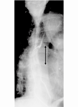
Fig. 1
Barium swallow showing ulcerative and localized tumor (black arrow) about 4 cm in length in the middle thoracic esophagus.
An aberrant right subclavian artery (ARSA) is classified as an anomaly of the aortic arch complex, and is regarded as one of the vascular rings, which sometimes causes compression of the trachea or esophagus [5]. The incidence of this anomaly in Japanese adults ranges from 0.15 to 1.6% with an average of about 0.5% [2]. Embryologically, in the normal individual, the proximal part of the right subclavian artery develops from the right 4th primitive aortic arch and the distal from the 7th intersegmental artery, but abnormal involution of the 4th right aortic arch causes persistence of the intersegmental artery which assumes a retroesophageal position [6].
This type of vascular anomaly always causes a non-recurrent inferior laryngeal nerve (NRILN) on the right side, because the 4th right aortic arch, where the right inferior laryngeal nerve generally recurs, disappears abnormally [5]. Therefore, when an ARSA is identified by means of CT, it can be predicted that the patient has a right NRILN. To our regret, we could not find the ARSA preoperatively in our case; it ran rightwards between the esophagus and the vertebral column (Fig. 2). Moreover, we did not identify a right NRILN during the operation. Had we also performed a cervical procedure, we would have discovered it in the right side of the neck, but we had decided not to dissect cervical lymph nodes before the operation because of his cardiac and respiratory dysfunction and absence of swelling of cervical lymph nodes.
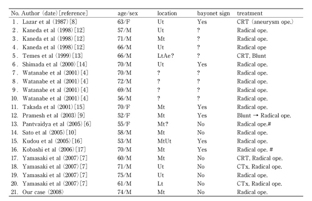
We investigated 12 case reports (21 patients) of esophageal cancer with an ARSA (including our case), using PubMed and Japana Centra Revuo Medicina (Table 1). Most of these cases were reported during the last 10 years and were also from Japan. The reason is probably that many patients have received esophagectomy for esophageal cancer in Japan and Japanese surgeons have often performed complete lymph node dissection along the bilateral recurrent nerves, and that they may find an ARSA or a NRILN with less difficulty than surgeons in other countries. Of the 21 patients, the average age was 65, and the male-female ratio 17 to 4. This is almost the same as in esophageal cancer patients without an ARSA. The tumors were located at the upper thoracic esophagus in 6 patients (Ut: 6), at the middle in 9 (Mt: 9), and lower in 2 (Lt: 2). There is a higher occurrence rate of esophageal cancer at Ut with an ARSA than without an ARSA. Compression of the esophagus and transit disturbance of food due to an ARSA may have an influence on the high rate of occurrence at Ut [7]. The esophageal filling defect due to an ARSA, which is seen in a barium swallow, is known as the bayonet sign [8]. Six patients had a bayonet sign, but only two had dysphagia resulting from esophageal compression due to an ARSA (dysphagia lusoria). According to Lazar et al, the patient received an operation of aneurysm of an ARSA, but her dysphagia persisted. Subsequently, she was diagnosed as esophageal cancer [8]. Because the symptom of dysphagia lusoria may be indistinguishable from that of a carcinoma, a careful evaluation should be performed to rule out other underlying esophageal abnormalities.
Esophagectomy was performed in 20 patients. Most of the patients underwent the thoracoabdominal procedure but 2 had blunt dissection. One of these two patients finally underwent the thoracoabdominal procedure too, because of a possible injury to an ARSA in the transhiatal procedure [9]. Two other patients experienced thoracoscopic surgery, and one of them had a major complication in that an ARSA together with esophagus were transected by a linear endoscopic stapler [6]. Careful handling in the operation is required to prevent an ARSA from rupturing.
In order to avoid injuring a right NRILN, Sato and co-workers proposed that the cervical and abdominal procedures should be performed before the thoracic procedure, when an ARSA is identified in the preoperative CT. Identification of a right NRILN before upper mediastinal lymphadenectomy was considered important in avoiding unexpected nerve injuries and relieving the operator from the stress that he could not identify a right NRILN but nevertheless should perform mediastinal lymph node dissection [10].
This vascular anomaly is often accompanied by an anomalous thoracic duct [11]. This is mentioned only in the report of Yamasaki et al [7]. The thoracic duct runs a normal retroesophageal course in the mediastinum until it is intercepted by an ARSA, when it is deflected towards the right instead of turning to the left as it usually does. Finally, it reaches the right venous angle, accompanying an ARSA [11]. Surgeons may as well know this anatomical fact for avoiding not only chylothorax following esophagectomy but also thoracic duct injury on the occasion of inserting a catheter into a right sublavian vein. In our case, the thoracic duct was identified and ligated in the mediastinum, but we did not ascertain where it terminated.
In conclusion, although cases of esophageal carcinoma with an ARSA are uncommon, similar reported cases have recently increased in number. The most important thing is not to overlook a right retroesophageal subclavian artery in the mediastinal CT preoperatively, and to confirm that an ARSA is associated with a right NRILN and a right-sided terminating thoracic duct.
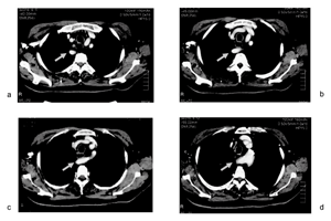
Fig. 2
Enhanced computed tomography showing a right retroesophageal subclavian artery (gray arrow), which arises from the aortic arch as its 4th branch (d), and runs rightwards between the esophagus and the vertebra (c, b) and upwards along the right side of the esophagus (a).
Table 1
Cases of esophageal cancer with an aberrant right subclavian artery
右鎖骨下動脈起始異常を伴う食道癌の1例を経験した。症例は74歳男性。食物つかえ感を主訴に来院。T3N3M0の胸部進行食道癌の診断のもと,右開胸開腹食道亜全摘,胃管による右胸腔内吻合術を行った。開胸時,食道背測の異常な右鎖骨下動脈を認めた。また右反回神経を同定できないまま,手術を終了した。術前評価で心肺機能の低下や頚部リンパ節腫脹のないことから,頚部リンパ節郭清は行わないことに決めていた。術後は肺炎以外の合併症もなく,軽快退院となった。
右鎖骨下動脈起始異常は大動脈弓部の先天性の奇形で,いわゆる血管輪の一つに数えられ,1%前後の頻度で発生する。この血管奇形はそれ自身が食道を圧迫し,dysphagia lusoriaを呈することもあるが,食道外科医にとって重要なことは,右反回神経が反回せず,頚部で迷走神経より直線的に分岐することである。また右静脈角に流入する胸管を合併することも多い。これらの解剖学的奇形は術前の造影CTによって診断が可能である。自戒を込めて,稀な血管異常を伴う食道癌の1例を報告した。
Department of Surgery, National Hospital Organization Chiba Medical Center, Chiba 260-8606.
森嶋友一,豊田康義,里見大介,青木靖雄,田沢洋一,白松一安,小林 純,鈴木一郎: 右鎖骨下動脈起始異常を伴う食道癌の1例.
国立病院機構千葉医療センター外科
Tel. 043-251-5311. Fax. 043-254-1349. E-mail: ym3300morishima@yahoo.co.jp
2008年2月13日受付,2008年6月16日受理.
