


Volume 84, Number 6
doi:10.20776/S03035476-84-6-P269
[Original Paper]
Hiroshi Tamai1), Masahiko Suzuki2), Yoshikazu Tsuneizumi3), Tadashi Tsukeoka4)
Scott A. Banks5), Hideshige Moriya1) and Kazuhisa Takahashi2)
(Received August 1, 2008, Accepted September 11, 2008)
The purpose of this study was to investigate the relation between long-term results and knee kinematics in the flat-on-flat CR-TKA. Sixteen knees were analyzed in the study. Fluoroscopic study of the step-up motion was done standing with the tibia at neutral, toe-in and toe-out positions relative to the femur in all implanted knees. Fluoroscopic analyses divided 16 joints into 2 kinematic groups: medial pivot, 8 and lateral pivot 8. The lateral pivot group showed anterior paradoxical movement in the toe-out position changing to the medial pivot pattern in the toe-in position, although the kinematics of the medial was not altered. In the 10-year follow-up, the average knee scores were 82.9±6.9 in cases of the lateral pivot group and 85.8±6.3 in cases of the medial pivot group. The average function scores were 59.4±14.6 in cases of the lateral pivot group and 57.0±11.6 in cases of the medial pivot group. The average flexion angles were 108.8±10.9° in case of the lateral pivot group 113.3±7.1° in case of the medial pivot group. There were no significant differences between the medial and lateral pivot groups in knee scores, function scores, and flexion angles. There were no cases of loosening or polyethylene failure.
total knee arthroplasty, knee kinematics, cruciate-retaining type, fluoroscopy, long-term results
Several fluoroscopic studies [1-13] after total knee arthroplasty (TKA) have revealed differences in kinematic patterns among cruciate-retaining (CR-), posterior stabilizing (PS-) and mobile-bearing total knee arthroplasties (TKA). Stiehl et al. [6] reported paradoxical anterior movement, condylar lift-off, and erratic screw home motion as risk factors related to abnormal wear characteristics in the flat-on-flat CR-TKA. Paradoxical anterior movement has been assumed to increase damage at the medial posterior edge of ultra high molecular weight polyethylene (UHMWPE), and to cause the polyethylene breakage in the short-term. Moreover, paradoxical anterior movement has the disadvantages of increasing the risk of patellofemoral instability due to lateralization of the tibial tubercle during deep flexion and decreasing maximum knee flexion because of reduced posterior femoral rollback of the lateral femoral condyle [12].
In 213 knees of in vivo fluoroscopic studies, Banks et al. [13] indicated that more constrained implant designs had less variability in the posterior translation of the femur, and the constraints promised successful total knee arthroplasties. In 1,027 knees of in vivo fluoroscopic studies, Dennis et al. [12] revealed that most fixed-bearing TKA designs rotated excessively beyond their design limits, and that mobile-bearing TKA designs have an advantage over fixed-bearing TKA designs to accommodate a wider range of axial rotation. Meanwhile, magnetic resonance studies showed medial pivot motion in healthy knee, and emphasized the importance of medial pivot motion in deep knee flexion [14-16]. Thus, some designs of total knee joints have been improved to induce medial pivot motion.
However, Ritter et al. [17] reported a 98.86% survival rate of 15-year follow-up in Anatomic Graduated Component (AGC) flat-on-flat CR-TKA. Further, there is no distinct evidence that total knee arthoplasties showing abnormal paradoxical motion have worse clinical results than those with medial pivot motion. Thus, the purpose of this study was to investigate the relation between long-term results and knee kinematics in the flat-on-flat CR-TKA.
Twenty-two patients (osteoarthritis; 14 knees in 11 subjects, rheumatoid arthritis; 15 knees in 11 subjects) underwent flat-on-flat CR-TKA in Chiba university hospital between April 1994 and July 1997. Fluoroscopic analyses were performed at 24.2 months (range, 12-42 months) average intervals between surgery and testing. Consent was obtained from all patients before surgery. At the 10-year follow-up, 1 case (2 knees) was lost, 7 cases (11knees) died from unrelated causes, and 16 knees in 14 subjects were analyzed in the study. Ten knees in 9 subjects had osteoarthritis and 6 knees in 5 subjects had rheumatoid arthritis. Two subjects had bilateral total knee arthroplasties. All 14 subjects were Japanese women with an average age of 63.8 years (range, 50-74 years). No cases of TKA after high tibial osteotomy and revision knee arthroplasty were included in the study. The clinical rating system of the Knee Society was applied for the evaluation of clinical results at the testing and 10 years after surgery.
All operations were performed by 1 senior surgeon (HM). The surgical technique involved approaching the knee through a medial parapatellar incision and then performing arthrotomy. After the anteroposterior dimensions of the distal femur were sized, the distal femoral cutting guide was aligned along a 0°-5° externally rotated axis from the posterior condylar axis, and the distal femur was cut. The proximal tibia was cut perpendicular to the axis of the tibial shaft. A posterior cruciate-retaining total knee prosthesis (Hi-Tech Knee IIR, Nakashima Medical Co. Ltd., Okayama, Japan) was implanted in each knee. The ligament balance at knee extension and flexion positions was confirmed by a manual examination, although no quantitative analysis was performed. The femoral, tibial, and patellar components were fixed without cement. The prosthesis design of the tibial insert was substantially flat in the sagittal and coronal planes. The femoral and tibial components were made of titanium alloy. The insert and patella were direct compression molded ultra high molecular weight polyethylene components.
In each subject, the implanted knee motion of a single step up a 17-cm stair was recorded by a lateral single plane fluoroscope (30 frames/sec) in 4 trials. Fluoroscopic study of the step-up motion was done again standing with the tibia at toe-in and toe-out positions relative to the femur in all implanted knees [20]. Using the model-matching technique [18] on computer systems, an in vivo 3-dimensional motion analysis of the implanted knee was performed. The anterior-posterior (A/P) translation and rotational patterns of the contact points of the medial and lateral condyles of the femoral components were analyzed on the tibial baseplate by examining the closing points between components. The midline was expressed as zero on the sagittal plane on the baseplate, and the contact points on the tibial baseplate were indicated. When the contact points moved anterior to the midline, the result was considered to be positive. Fluoroscopic data with the tibia at a position neutral to the femur were used to determine the centers of rotation. The lines connecting the medial and lateral contact points of each angle were plotted in each knee and the average centers of rotation were obtained. The cases in which the average centers were located between 0% and 100% at the lateral/medial center of rotation were defined as the medial pivot group. Similarly, the average centers of the lateral pivot group were between -100% and 0% for the lateral/medial center of rotation.
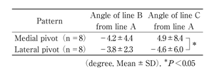
Computed tomography (CT) was performed on all implanted knees in the extended, supine position after the fluoroscopic study. In CT images, line A was drawn between the medial epicondyle and the lateral epicondyle of the femur (clinical epicondylar line). Line B was the tangent line of the anterior ridge of the pegs on the femoral component. In this design of the femoral component, line B was parallel to the posterior condylar line. Line C was the tangent line of the posterior ridge of the tibial baseplate (Fig. 1). The angle of line B from line A and the angle of line C from line A were measured in all implanted knees. When lines A and C were externally rotated from line B, the angles were expressed as positive.
Varus and valgus stress tests and a posterior drawer test were performed with 150N using a Telos® arthrometer (Telos, Medizinishch-Techniche GmbH, Griesheim, Germany). Posterior stability was evaluated by the midpoint method [21], in which the proportion of the distance between the anterior tip of tibia and the average contact points to the sagittal tibial diameter is measured. A ratio under 45% is suggestive of posterior instability .
Student's t test and Mann-Whitney rank sum test were used to compare means of the clinical and kinematic range data (P<0.05). Two-way repeated measure analysis of variance with post hoc pair-wise (Tukey's) comparisons were used to compare kinematic patterns over the range of flexion. The significance level was set at 0.05.
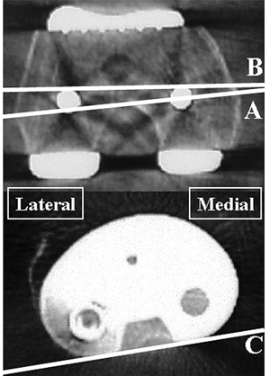
Fig. 1
Measurement of rotational alignment.
The angle of line B to line A. and the angle of line C to line A were measured in the extended, supine position. Line A, clinical epicondylar line; line B, the tangent line of the anterior ridge of the pegs on the femoral component; line C, the tangent line of the posterior ridge of the tibial baseplate.
At the testing, the average knee scores were 89.4±4.6 for all cases, 90.1±3.4 for cases of osteoarthritis (OA), and 88.4±5.6 for cases of rheumatoid arthritis (RA). The average function scores were 76.0±10.4 for all cases, 77.1±10.4 for cases of OA, and 74.4±5.8 for cases of RA. There were no significant differences between OA and RA in knee scores and function scores.
The analyses of the centers of rotation classified all knees into the medial pivot (MP) group or the lateral pivot (LP) group (Fig. 2).
(1) Medial pivot group (8 joints) : The lateral condyle moved posteriorly at knee flexion with a medial pivot. The medial condyle also moved posteriorly, but the distance of translation was smaller than that of the lateral condyle (P<0.05).
(2) Lateral pivot group (8 joints) : The medial condyle moved anteriorly at knee flexion with a lateral pivot. The lateral condyle also moved anteriorly, but the distance of translation was smaller than that of the medial condyle (P<0.05). This motion was considered to be paradoxical movement.
The medial pivot had no change in kinematic patterns between toe-in and toe-out positions, but the lateral pivot group showed the lateral pivot pattern in the toe-out position, then changed to the medial pivot pattern in the toe-in position (Fig. 3).
In valgus and varus stress tests in knee extension, the joint angles of the varus stress test were 2.6°+3.5° in the medial pivot group and 2.9°+1.4° in the lateral pivot group, and the joint angles of valgus stress test were 4.4°+2.4° in the medial pivot group and 3.4°+1.3° in the lateral pivot group (Table 1). There were no significant differences between the varus stress and valgus stress tests in these two groups. The posterior drawer tests showed no posterior instability in both medial and lateral pivot groups.
Computed tomography scans showed mean angles of the epicondylar line of the femoral condyle against the tangential line of the tibial tray to be 4.9°+8.4° in the medial pivot group and -4.6°+6.0° in the lateral pivot group, which were statistically significant (P<0.05) (Table 2). There were no significant differences between the kinematic patterns and the externally rotated setting of the femoral component, although the distal femoral cutting, which was aligned along the 0°-5° externally rotated axis from the posterior condylar axis, caused variability in the angle of line B from line A.
At the 10-year follow-up, the MP group had osteoarthritis in 5 knees and rheumatoid arthritis in 3 knees. The LP group had osteoarthritis in 5 knees and rheumatoid arthritis in 3 knees. The average knee scores were 84.3±6.6 in all cases, 82.9±6.9 in cases of LP, and 85.8±6.3 in cases of MP. The average function scores were 58.2±12.9 in all cases, 59.4±14.6 in cases of LP, and 57.0±11.6 in cases of MP. The average flexion angles were 111.7±9.1°. in all cases, 108.8±10.9° in case of LP, 113.3±7.1° in case of MP. There were no significant differences between the medial pivot group and the lateral pivot group in knee scores, function scores, and flexion angles. There were no cases of loosening or polyethylene failure.
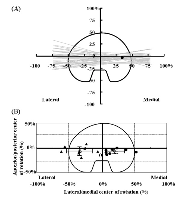
Fig. 2
The centers of rotation in medial and lateral pivot cases.
(A) : The fluoroscopic study in a 71-year-old woman represents the medial pivot pattern due to the average center of rotation at the medial side of the tibial tray. Gray lines show the instantaneous flexion-extension axis of the femur projected onto the tibial baseplate for stair activities between 0° and 90° of knee flexion. ●: the average center of rotation.
(B) : In 16 knees, the average centers of rotation were divided into medial and lateral pivot patterns. ●: the average centers of rotation in each medial pivot patient, ○: the average center of rotation for the entire medial pivot group, ▲: the average centers of rotation in each lateral pivot patient, △: the average center of rotation for the entire lateral pivot group. The length of each arm of the black cross indicates the standard deviation of the mean.
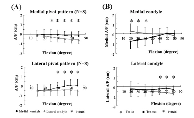
Fig. 3
The knee kinematics in flat-on-flat CR-TKA.
(A) The kinematic patterns of the 2 groups.
(B) The change in kinematics in the lateral pivot group.
Table 1
Results of stress test.
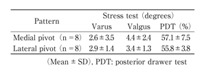
Table 2
Results of rotational alignment.
Several studies have revealed knee kinematics using MRI and RSA [19,20,22]. Hill et al. [20] and Karholm et al. [22] reported the change of contact points based on tibial rotation in the loaded natural knee. In particular, Hill et al. [20] showed that the medial pivot movement in neutral and internal tibial rotations changed to the uniaxial hinge in external rotation. Similarly, the lateral pivot group in the study showing a paradoxical movement in the toe-out position dramatically changed to the medial pivot patterns in the toe-in position.
In a computed tomography study, cases in which the rotational alignment of the femoral component did not match with their epicondylar line did not always indicate abnormal kinematics in CR-TKA. The lateral pivot group showed external rotation of its epicondylar line against the tangential line of the tibial tray. The relative setting of components in femur and tibia is assumed to be one of the factors affecting knee knematics. Nozaki et al. [23] reported that kinematic patterns were different among surgeons in unconstrained CR-TKA. Nevertheless, surgical techniques regarding the knee kinematics were not mentioned. Implant design, surgical technique of TKA, and anatomic variance are considered to relate to axial rotation [12]. However, definite conclusions on surgical technique have not yet been reached.
A series of fluoroscopic studies [1-13,24,25] has been performed to find ways to improve range of motion, durability of implant, and long-term results. Prosthetic designs for CR-TKA substantially showed variable kinematic patterns and anterior paradoxical movement [10-13]. In prosthetic designs for PS-TKA, the engineer-intended and definitive kinematics such as medial pivot pattern could be observed [6,10-13]. Banks et al. [13] revealed that 39 percent (50 of 129 knees) of CR-TKA had medial centers of rotation during a stair-climbing activity. Dennis et al. [12] showed a huge variability in the kinematics of CR-TKA. Thus, 129 of 163 knees (79%) had a medial rotation pattern from full extension to 90° of knee flexion, whereas 130 of 163 knees (80%) had a reverse rotation pattern during at least 1 flexion increment. In the study, 50% (8 of 16 knees) of CR-TKA had a constant medial center of rotation, and the remaining 50 percent (8 of 16 knees) showed variability in response to toe position.
Todo et al. [26] reported that 2-dimensional sliding fatigue testing increased crack formation and propagation in UHMWPE more than single sliding fatigue testing, and the least variability of knee kinematics was of value in decreasing polyethylene wear. However, a series of AGC flat-on-flat CR-TKA maintained good long-term results in patients with osteoarthritis and rheumatoid arthritis [17,27]. In the study, both medial and lateral pivot groups showed good results for knee score and knee flexion angle. The function score decreased to 58 points because of unrelated causes such as cardiac disease, femoral neck fracture, and spinal canal stenosis. Medial pivot motion might be meaningful in inducing the additional posterior translations of both femoral condyles on flexion beyond 120° [28].
There are some limitations to the current study. Patients with osteoarthritis and patients with rheumatoid arthritis were enrolled in this study. The ratios of rheumatoid arthritis patients were same in both groups. Kinematic patterns might change in the course of time. Knee stability and alignment in the knee score were similar at the testing and 10-year follow-up, and this result showed less possibility of a change in knee kinematics.
In this study, the medial pivot group and the lateral pivot group at 10-year follow-up had good clinical results even for flat-on-flat cruciate-retaining total knee arthroplasties. However, the number of cases was limited and larger scale studies are required in the future.
人工膝関節術後の膝関節透視下動作解析による研究では,anterior paradoxical movementを示す人工膝関節はポリエチレンの摩耗や破損を加速し長期成績が不良となると報告されている。本研究では後十字靱帯温存型人工膝関節手術後の患者が階段を昇るときの運動様式を評価して10年後の臨床成績と比較検討した。22症例29関節で動作解析を行ったが,10年のfollow-upが可能だったのは14症例16関節だった。16関節は透視下動作解析により2グループに分かれ,medial pivot groupが8関節,lateral pivot (anterior paradoxical movement) groupが8関節だった。10年経過では,knee scoreの平均はlateral pivotグループで82.9±6.9点,medial pivotグループで85.8±6.3点だった。Function scoreの平均はlateral pivotグループで59.4±14.6点,medial pivotグループで57.0±11.6だった。平均屈曲角度はlateral pivotグループで108.8±10.9°,medial pivotグループで113.3±7.1°だった。Medial pivotグループとlateral pivotグループではknee score,function score,屈曲角度に有意差はなく,人工関節の緩みやポリエチレンの破壊により再置換術となった症例もないことから,後十字靱帯温存型人工膝関節の運動様式は長期成績に影響を与えないと考えられた。
玉井 浩1),鈴木昌彦2),常泉吉一3),付岡 正4),Scott A, Banks5),守屋秀繁1),高橋和久2): 後十字靱帯温存型人工膝関節の運動様式は長期成績に影響を与えない.
1) 鹿島労災病院整形外科, 2) 千葉大学大学院医学研究院整形外科学, 3) 上都賀総合病院整形外科, 4) 金沢病院整形外科, 5) フロリダ大学整形外科
Tel. 043-226-2117. Fax. 043-226-2116. E-mail: masahiko@faculty.chiba-u.jp
2008年8月1日受付,2008年9月11日受理.
