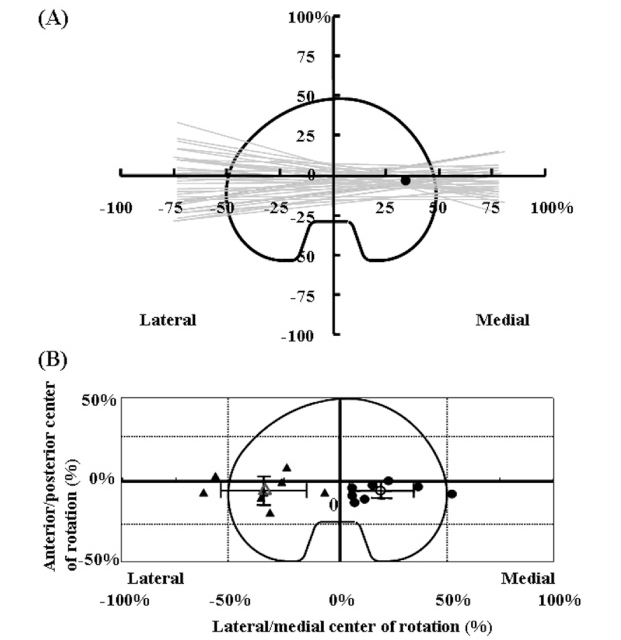
Fig. 2
The centers of rotation in medial and lateral pivot cases.
(A) : The fluoroscopic study in a 71-year-old woman represents the medial pivot pattern due to the average center of rotation at the medial side of the tibial tray. Gray lines show the instantaneous flexion-extension axis of the femur projected onto the tibial baseplate for stair activities between 0° and 90° of knee flexion. ●: the average center of rotation.
(B) : In 16 knees, the average centers of rotation were divided into medial and lateral pivot patterns. ●: the average centers of rotation in each medial pivot patient, ○: the average center of rotation for the entire medial pivot group, ▲: the average centers of rotation in each lateral pivot patient, △: the average center of rotation for the entire lateral pivot group. The length of each arm of the black cross indicates the standard deviation of the mean.