


Volume 84, Number 6
doi:10.20776/S03035476-84-6-P277
[Case Report]
Suguru Shirato1,3), Katsuhiro Hanawa1), Hiroshi Nagata2)
Emiko Adachi-Usami1) and Shuichi Yamamoto3)
(Received June 17, 2008, Accepted July 14, 2008)
Purpose. To report a case of branch retinal artery occlusion including the ophthalmic artery posterior to the optic disk following cataract surgery in a patient with bilateral internal carotid artery stenosis.
Case report. A 75-year-old woman underwent phacoemulsification and intraocular lens implantation in her right eye. The surgery was uneventful. A month later, her vision of the operated eye decreased and an inferior altitudinal visual field defect was found, and ophthalmoscopy revealed two superior branch retinal artery occlusions. Fluorescein angiography showed extremely delayed infusion of the two arteries and the vessels in the disk. The single flash, rod-cone electroretinogram had a negative-type pattern. Magnetic resonance imaging angiography and Doppler echography demonstrated bilateral carotid artery stenosis. The avascular area of the retina was photocoagulated, and aspirin was prescribed. No further ocular complications occurred.
Cataract surgery, Doppler echography, MRA, Ocular Ischemic Syndrome, Carotid artery stenosis
Even though the surgical techniques and instrumentation for cataract surgery have significantly improved, complications following successful surgery still occur. We report a rare case of ocular ischemic syndrome (OIS) caused by a stenosis of the carotid artery that developed within a month after cataract surgery.
A 75-year-old woman presented with decreased vision of 20/32 OD and 20/25 OS. Informed consent was obtained from the patient. Her fundi were normal except for slight sclerotic changes of the retinal vessels. She was diagnosed with cataract and was followed for 4 years. When her visual acuity decreased to 20/50 in both eyes, phacoemulsification with intraocular lens (Acrysof, Alcon) implantation was performed in her right eye.
A day after surgery, her vision had improved to 20/20 in the right eye, but on the next appointment a month later, the visual acuity had decreased to 20/100. Her right fundus revealed white sheathing of the superior retinal arteries, with soft exudates and hemorrhages near the disc. The disc was slightly pale (Fig. 1, left). There were no abnormal vessels on the iris and angle, and the intraocular pressure was 14 mm Hg in both eyes. Goldmann and Humphrey automated perimetry demonstrated an inferior altitudinal visual defect. The b-wave of the single bright flash ERG of the right eye was greatly depressed while the a-wave was of normal size, i.e., a negative-type ERG. The b- to a-wave ratio was 0.8, in the right and 1.0 in the left. The oscillatory potentials were not recordable in both eyes (Fig. 2). Pattern visually evoked cortical potentials were non-recordable with stimulation of the right eye.
Fluorescein angiography showed delayed perfusion in the two superior arteries and staining of the disc. There were avascular areas in the superior peripheral retina (Fig. 1, right), and the avascular areas were photocoagulated with a laser.
MRI and MRA of the brain, MRA of the carotid artery, and Doppler echography were planned to try to find the cause of the branch retinal artery occlusion and ischemic optic neuropathy. After explaining the purpose and procedures to be performed, an informed consent was obtained. The procedures were conducted to conform to the tenets of the Declaration of Helsinki.
These tests showed marked stenosis of both internal carotid arteries with the degree of stenosis approximately the same in both arteries (Fig. 3). Multiple high-intensity lesions involving the white matter of both hemispheres (Fig. 4) were detected but she had no symptoms. Aspirin was prescribed and her vision has varied between 20/63 and 20/32 in both eye during the 4 month follow-up period.
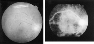
Fig. 1
Fundus photograph (A) and fluorescein angiogram (B) of the right eye showing delayed dye infusion into the temporal and nasal superior retinal arteries and in the optic disc.
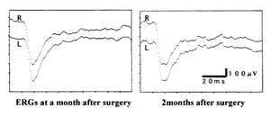
Fig. 2
Single flash maximal ERG demonstrating a negative-type pattern with greatly reduced oscillatory potentials. The response became more abnormal with time. A: one month after surgery, B: two months after surgery.
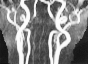
Fig. 3
MR angiography demonstrating stenosis at the siphon of the right carotid artery.
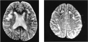
Fig. 4
T2 MR imaging of the brain demonstrating multifocal ischemic lesions in both hemispheres.
It is somewhat difficult to explain the relationship between the development of OIS and cataract surgery, and whether the ocular ischemic changes were triggered by the cataract surgery. It could have happened by chance but the occlusion of the retinal artery and the ophthalmic artery within the optic disc was in the eye that underwent cataract surgery and occurred within a month after the surgery. Unfortunately, the exact onset of the occlusion could not be determined because the patient was not aware of the decreased vision in that eye. It was presumed that the OIS occurred shortly after surgery, as the fundus appearance at a month later already revealed silver wire arteries.
It is highly likely that the stenosis of the internal carotid artery was present before the cataract surgery, but the stenosis was not suspected because she had no symptoms or any signs of retinal ischemia, although she had systemic hypertension. It should be remembered that the carotid stenosis was bilateral and approximately equal, and the OIS developed in the eye that underwent cataract surgery. Accordingly, intraocular perfusion pressure caused by surgery could be responsible for the OIS, too.
A Medline search extracted only two cases of retinal artery occlusion following cataract surgery. The first case [1] was a 75-year-old man who developed neovascular glaucoma 3 months after unilateral extracapsular cataract extraction with intraocular lens implantation. Carotid angiography demonstrated occlusion of the ipsilateral right internal carotid artery, and a steal phenomenon from the right ophthalmic artery. The second case was reported by Chan et al [2] who retrospectively reviewed 142 eyes with posterior capsule rupture during cataract surgery. They found one case with a central retinal artery occlusion following cataract surgery.
Jurowski et al [3] measured the nitric oxide (NO) level in the aqueous humor of rabbits and found an increased NO level after lensectomy, although the use of phacoemulusification with the insertion of a foldable intraocular lens, as we did, caused only a slight release of NO compared to other surgical techniques with different intraocular lenses. This is relevant to our patient because NO plays an important role in regulating local blood flow [4,5] and NO also mediates the neurotoxic action of glutamate that is responsible for the ischemic retinal injury. In addition, elevated NO has been found in eyes with CRAO. [6,7] It is possible that excessive NO production may have contributed to the pathological developments in the anterior chamber and retina, such as extensive vessel dilation, changes in vessel permeability, and a breakdown of the blood-retinal barrier.
In conclusion, our findings suggest that cataract surgery may be a risk factor for the development of ocular ischemic syndrome. We recommend a careful examination of OIS after intraocular surgery in elderly patients with systemic vascular problems.
The authors thank for Professor Duco Hamasaki, Bascom Palmer Eye Institute, for editing the manuscript.
75歳男性,両内頚動脈狭窄,右眼白内障,眼内レンズ挿入術施行1ヵ月後,術眼に眼虚血症候群が生じた症例を報告した。両上耳側,鼻側の2本の中心動脈の閉塞が眼底像で認められ,蛍光眼底造影にてこの2本の細動脈の著しい血流遅延および周辺網膜に無血管野が示された。網膜電図は陰性型を呈した。MRIにて両側の内頚動脈の狭窄,頚部のドップラーエコーグラムでも両内頚動脈の血流遅延を認めた。右眼の無血管野にレーザー光凝固を施行し,その後視機能の増悪はみられていない。内頚動脈狭窄がある場合,白内障手術が眼虚血症候群を発症させる起因となることについて考按した。
白戸 勝1,3),塙 勝博1),永田博史2),安達惠美子1),山本修一3): 白内障手術後に眼虚血症候群を発症した1例.
1) 山王病院感覚器病センター眼科
2) 山王病院感覚器病センター耳鼻咽喉科
3) 千葉大学大学院医学研究院眼科学
Tel & Fax. 043-421-1772. E-mail: eadachi-2003@coral.ocn.ne.jp
2008年6月17日受付,2008年7月14日受理.
