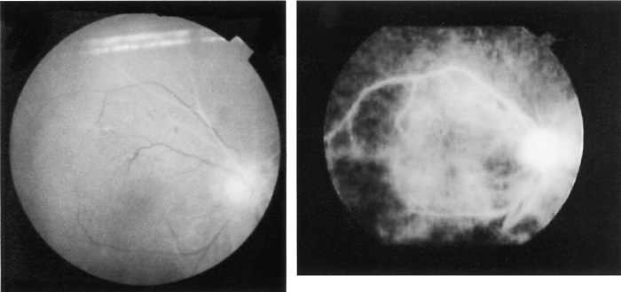
Fig. 1
Fundus photograph (A) and fluorescein angiogram (B) of the right eye showing delayed dye infusion into the temporal and nasal superior retinal arteries and in the optic disc.
Fig. 1
Fundus photograph (A) and fluorescein angiogram (B) of the right eye showing delayed dye infusion into the temporal and nasal superior retinal arteries and in the optic disc.