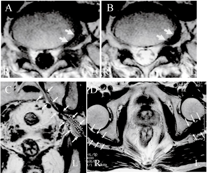
Fig. 1
Axial spin-echo T1- and T2-weighted magnetic resonance imaging (MRI) revealed left extraforaminal disc herniation at L5/S1 level before initial surgery (A and B; arrows). Frontal view of MRI revealed the left sciatic nerve (C; arrows), and axial view of MRI revealed piriformis muscle (D; arrows). Dotted lines indicated bifurcation of the sciatic nerve (C). The piriformis muscle on the left side was thicker than that on the right side R: right side, L: left side.