


Volume 86, Number 6
doi:10.20776/S03035476-86-6-P219
[Original Paper]
Tomoaki Tatsumi, Shuichi Yamamoto and Ichiro Shimoyama1)
(Received June 18, 2010, Accepted July 9, 2010)
【Purpose】To find automated diagnosis of retinopathy, the matched filters (Chaudhuri et al, 1989) were studied for retinopathy. The filters had been reported for a film-based fundus image of 2.5x105 pixels, and the image needed to be converted into a digital file with an image scanner. Now, direct digital images are available with more precise images than 107 pixels.
【Methods and Subjects】Non compressed digital images were taken with a non mydriasis-type fundus camera, and images of green component were used. Fluorescein angiography was done for retinopathy, and the images were compared to the matched filters. Subjects were 11 normal volunteers and 25 patients with typical diabetic retinopathy, 7 patients with branch retinal vein occlusion, 18 patients with age-related macular degeneration, respectively.
【Results】Fine retinal blood vessels, microaneurysms, neovascularization, dotted retinal bleedings and exudates were emphasized for diabetic retinopathy, but non-perfusion areas in fluorescein angiograms could not be made clear with the filters.
【Conclusion】The filters would be helpful for diagnosis of retinopathy, but the process could not alternate with fluorescein angiography. The filters were not good for the diseases of transparent part such as cataract or vitreous hemorrhage.
matched filters, fundus camera, retinopathy, screening
Certain systemic diseases are productive of retinopathy as diabetes, hypertension, or atherosclerosis. The retinopathy threatens the patients with blindness, so those diseases impact on the quality of life. Population with diabetes was 2.8% in 2000 and estimated to rise to 4.4% in 2030[1], therefore, the screening of ocular fundus is becoming very important. Fundus images could be observed directly to detect retinopathy as retinal bleeding or glaucoma. The images were taken easily as photographs, so they are appropriate for a screening examination.
Fundus photographs, taken in outpatients of internal medicine or health screening, are taken for medical disorders such as hypertension or diabetes in Japan. The diagnosis at the first step is done by a physician, not an ophthalmologist in many cases, so there is any chance of misdiagnosis for the images. Automated diagnosis of fundus photographs will be helpful for physicians. With abnormal findings by the automated diagnosis they can refer to ophthalmologists for further examination. In checking the screening photographs by ophthalmologists, they have much time for retinopathy with the reports of the automated diagnosis.
Chaudhuri et al. had reported the matched filters for fundus photographs in 1989 as filters to emphasize vessel, they studied an algorithm to detect the edges of retinal blood vessels. They studied the algorithm for the normal fundus images of around 2.5x105 pixels and reported it useful to detect blood vessels[2]. They did not operate the algorithm on line in real time at the time, the fundus images were taken by a camera to a film and the film developed and enlarged on a sheet of photographic paper, then the images on the paper were converted to a digital file by an image scanner, then the file was operated with the algorithm. And, Hoover et al., Lowell et al. improved the algorithm[3,4]. Now, a digital file can be directly obtained from a digital camera of a fundus camera with higher resolution than 1.0x107 pixels, and the file is ready for operating them with the algorithm using a conventional personal computer.
In cases of progressive diabetic retinopathy, microaneuryms, occlusion of retinal blood vessels, ischemic areas, neovascularization and exudates occur frequently. The retinopathy is irreversible, so early detection is critical to stop the progression. Fluorescein angiography can easily detect hyper-perfusion areas, but, the angiography is used to doing at an ophthalmologist and anaphylactic shock occurred accidentally.
Spencer et al. reported a morphological transformation to segment microaneurysms from fluorescein angiograms[5], and the method was improved[6,7]. Hipwell et al. reported a method of red-free images for the microaneurysms[8]. Neural network had been reported for diabetic retinopathy or vessel abnormality[9-11].
In cases of age-related macular degeneration, hemorrhage and choroidal neovascularization occur at macula. The abnormal findings are obvious in advanced stage, but they might be difficult to detect them in early stage. Poor studies have been reported to detect abnormal blood vessels or hemorrhages for age-related macular degeneration.
With those diseases, automated detection of abnormal vessels and/or exudates should be helpful and useful for not only physicians, but also ophthalmologists who were checking the large number of images from fundus screening.
We studied the matched filters with high-resolution fundus digital images for typical retinopathies.
Subjects were 49 eyes of 25 patients (57.3±10.2 y/o) with typical diabetic retinopathy, 7 eyes of 7 patients (66.0±9.7 y/o) with branch retinal vein occlusion, 21 eyes of 18 patients (69.1±11.2 y/o) with age-related macular degeneration and 11 eyes of 11 volunteers with normal fundus. Informed consent was obtained from all subjects.
A digital camera (D80®, Nikon, Japan) was attached to a non mydriasis-type fundus camera (TRC-NW6S®, Topcon, Japan) to take images as non-compressed digital files. Fluorescein angiography wad done for the subjects except for a normal subject, and the fluorescein angiograms were compared with images by the matched filters. Fluorescein angiograms were taken with a fundus camera (TRC-50IX®, Topcon, Japan) with a digital camera (D1x®, Nikon, Japan) after intravenous injection of a fluorescent medium (FLUORECITE®, Alcon Japan Ltd., Tokyo, Japan) 300mg, and saved as the joint photographic expert group format (jpg or jpeg).
Non compressed files were converted to the tagged image file format (tif or tiff), and green components were used for discussion. The algorithm of the matched filters was below, following Chaudhuri et al.[2]. The retinal blood vessel does not have most of ideal step edge in the gray-level profiles along directions perpendicular to their length. Although the intensity profile varies by a small amount from vessel to vessel, it may be approximated by a Gaussian curve f (x, y) = A {1-k exp (-d2/2σ2)}. Where d is the perpendicular distance between the point (x, y) and the straight line passing through the center of the blood vessel in a direction along its length, σ defines the spread of the intensity profile, A is the gray-level intensity of the local background, and k is a measure of reflectance of the blood vessel relative to its neighborhood.
s0( t ): output signal
H( f ): filter
S( f ): Fourier transform of s( t )
η( f ): noise spectrum
S( t ): input signal
The concept of the matched filters is extended to two dimensional images, the process was performed every 15 degree in 12 directions and at each pixel only the maximum of their responses was retained.
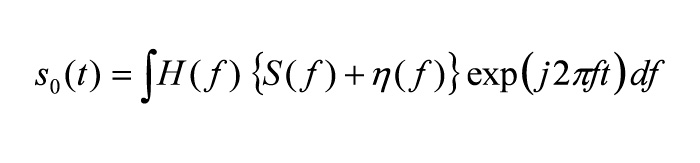
Fig 1(Fig. 1) showed an example for a normal volunteer; an upper image was an original image taken by a camera, 3 images in the middle raw were 3 components of red (R), green (G) and blue (B), 3 images in the lower raw were operated images with the matched filters, corresponding to the 3 components. Blood vessels were markedly noted in the image of the green component, and were remarked for the operated image of the green component.
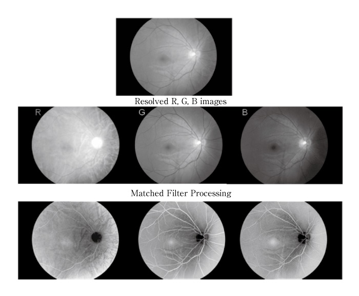
Fig. 1
Example images for a normal volunteer; the upper image was the original image, 3 images at the middle raw were 3 components of red (R), green (G) and blue (B), 3 images at the lower raw were operated images corresponding to the above 3 components with the matched filters.
Fig 2(Fig. 2) showed an example for non-proliferative diabetic retinopathy; the left image was original, the middle image was obtained with fluorescein angiography, and the right image was operated image by the matched filters. Many microaneurysms were noted in 3 images, microaneurysms were the most noted in the fluorescein angiography. Many dotted retinal bleedings were noted in the original and operated images, but they were represented as hypo-perfusion areas in the fluorescein angiography.
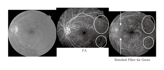
Fig. 2
Example images for non-proliferative diabetic retinopathy; the left image was the original image, the middle image was for fluorescein angiogram (FA), and the right image was for the matched filters. Microaneurysms were noted for 3 images. The white circles showed non-perfusion areas, and the arrows showed nerve fivers emphasized.
Fig 3(Fig. 3) showed proliferative diabetic retinopathy, the left image was original, the middle image was for the fluorescein angiography, and the right image was operated image by the matched filters. Retinal bleedings were noted in the original image and they were represented as hypo-perfusion areas in the fluorescein angiogram. Neovascularization was noted in the fluorescein angiogram.
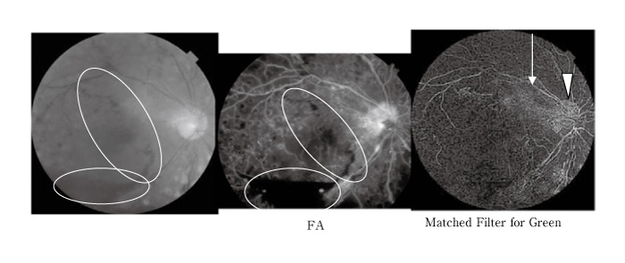
Fig. 3
Example images for proliferative diabetic retinopathy; the left image was the original image, the middle image was for fluorescein angiogram (FA), and the right image was for the matched filters. The ellipses showed vitreous hemorrhage, the arrows showed linear structure for proliferative tissue, and the arrow head showed neovascularization and proliferative tissue.
Fig 4(Fig. 4) showed old branch retinal vein occlusion; the left image was original, the middle image was for fluorescein angiogram, and the right image for the matched filters. The white arrow showed the venous occlusion, a whitish streak was noted in the original image, and it was obscure in the fluorescein angiography and by the matched filters. Neovascularization was noted in the white circle adjacent to the macula in the fluorescein angiography and the image by the matched filters. Abnormal clusters of white spots were noted surrounding the circle in the image by the matched filters, which was not noted in the fluorescein angiography, and noted as soft exudates in the original image.
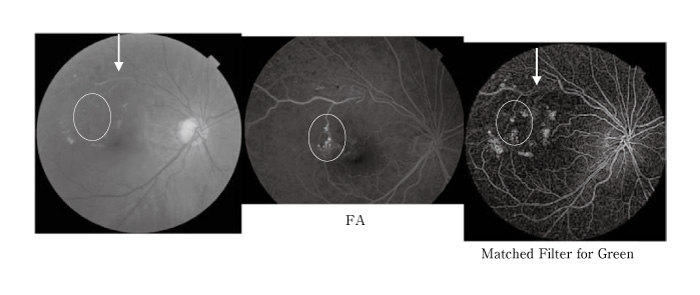
Fig. 4
Example images for old branch retinal vein occlusion; the left image was original, the middle image was for fluorescein angiogram (FA), and the right image for the matched filters. The arrows showed occluded vein, and the circles showed aneurysms. Exudates were noted for the original and the matched filters.
Fig 5(Fig. 5)showed age-related macular degeneration; the left image was original, the middle image was for fluorescein angiogram, and the right image for the matched filters. The original image showed hard exudates and hemorrhage, and the fluorescein angiograms showed leakage. The matched filters emphasized the hard exudates, but could not emphasize the hemorrhage on macula. The fluorescein leakage could not be noted for the matched filters.
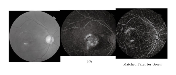
Fig. 5
Example images for age-related macular degeneration; the left image was original, the middle image was for fluorescein angiogram (FA), and the right image for the matched filters. The exudates on the original image were emphasized for the matched filter, but fluorescien leakage for FA did not detected at all for the matched filters.
The matched filters could emphasize fine vessels, and neovascularization and microaneurysms could be emphasized for diabetic retinopathy, branch retinal vein occlusion, age-related macular degeneration. The matched filters, theoretically, were restricted to emphasize images; original images with poor signals could not be emphasized, so, the diseases were not indicated for the filters of transparent parts like as cataract or retinal- or vitreous- bleedings. The images except for edges of vessels could be emphasized like as hard exudates, which was good to fundus screening. The findings were easy to differentiate between 2 pathogeneses by comparing images with the original color image. And the filters were only mathematical operation, the filters could not detect the hemodynamics as the fluorescein angiography.
A red-free filter has been used to observe fundus vessels, and the green component was good to proceed with the matched filters (Fig. 1-G). The red component was good to observe deep choroids plexuses (Fig. 1-R). The blue component was good to observe surface nerve fiber layer (Fig. 1-B).
The case with non-proliferative diabetic retinopathy showed lots of dotted hemorrhages and microaneurysms in the original, the fluorescein angiogram and the matched filtered images (Fig. 2). Nerve fibers were emphasized in the image by the matched filters (white arrows). Non-perfusion area found in peripheral area becomes slightly dark (white circles). It was thought that blood circulation was intercepted, and the retinal thicknesses decreased, and reflections of the light decreased. However the finding with the matched filters was not clear, the hemodynamics in fluorescein angiography could not be estimated with the matched filters.
The case with proliferative diabetic retinopathy showed vitreous hemorrhage for the original and the fluorescein angiogram in the circles, but poor visualization for the image by the matched filters (Fig. 3). Information beneath the hemorrhage had not been detected on the original image, so the matched filters were in vain. However, neovascularization and proliferative tissue on and around the optic disc were emphasized markedly by the matched filters (the white arrow head). The meshwork structure on the optic disc was thought as neovascularization for the matched filters image. And the linear structure extending upward along arcade vessel from optic disc was thought to contain proliferative tissue (the white arrow).
The case with old branch retinal vein occlusion showed the white sheathed blood vessel in the original and the matched filters images (the white arrow head in Fig. 4). Different emphasized pattern was noted from normal vessel and only the vascular walls were emphasized (the white arrow). An aneurysm-like shape was noted for the matched filters and the fluorescein angiogram (the white circle), but it was hard exudates for the original image.
The case with age-related macular degeneration showed exudates surrounding the macula for the original image, and the fluorescein leaked much over the macula in the fluorescein angiogram, but the exudates were emphasized and the leakage of exudates could not be detected for the matched filters (Fig. 5).
In conclusion, clinical application of the matched filters provided us much information with a digital fundus camera. Fine vessels such as neovascularization and microaneurysms, exudates and dotted hemorrhage were emphasized. But, the filters were not good for cases with opacity of the transparent body. The filters could not evaluate the dynamics of blood flow, and the filters could not take the place of fluorescein angiography.
The authors thank Professor Yoichi Miyake (Frontier Medical Engineering, Chiba Univ.) for his useful suggestion.
【目的】網膜像の血管強調を目的として,Matched Filter法が開発された(Chaudhuri et al, 1989)。Chaudhuriらの報告では,2.5x105画素数の画像に,手動でイメージスキャナーを使用してデジタル化したが,現在では107画素数以上の高解像度画像を直接処理可能になった。今回,正常眼底および網膜症の高解像度カラー画像に対してこのMatched Filter法を用いて解析をおこなった。
【方法】無散瞳型眼底カメラに装着したデジタルカメラでカラー眼底写真を非圧縮撮影し,非圧縮保存し,緑成分の画像をMatched Filter法により処理した。正常眼底11眼,典型的な糖尿病網膜症7眼,網膜静脈分枝閉塞症21眼,加齢黄斑変性21眼を対象とし,研究の趣旨を説明し同意後解析した。網膜症に対しては蛍光眼底造影検査を施行し,その画像をMatched Filter法画像と比較検討した。
【結果】Matched Filter法処理により細い網膜血管が強調された。糖尿病性網膜症では網膜最小血管瘤と新生血管が強調された。しかし,蛍光眼底造影により得られた無潅流領域はこの画像処理により明確にすることができなかった。
【結論】Matched Filter法処理により網膜最小血管瘤や新生血管のような異常血管を強調することが可能であり,網膜症の診断に有用である可能性が示唆された。しかし,蛍光眼底造影検査の代わりになるものではない。さらに白内障や硝子体出血のような中間透光体に混濁があり画像撮影不良な疾患には限界があるが,スクリーニングの自動判定に有用であることが結論された。
Department Ophthalmology and Visual Science, Graduate School of Medicine, Chiba University, Chiba 260-8670.
1) Section for Human Neurophysiology, Research Center for Frontier Medical Engineering, Chiba University, Chiba 263-8522.
辰巳智章,山本修一,下山一郎1): 高解像度眼底画像に対するMatched Filter処理の臨床応用.
千葉大学大学院医学研究院眼科学講座
1) 千葉大学フロンティアメディカル工学研究開発センター
Tel. 043-222-7171. Fax. 043-226-2281. E-mail: newyear98mt@yahoo.co.jp
2010年6月18日受付,2010年7月9日受理.
