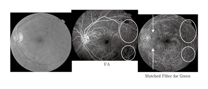
Fig. 2
Example images for non-proliferative diabetic retinopathy; the left image was the original image, the middle image was for fluorescein angiogram (FA), and the right image was for the matched filters. Microaneurysms were noted for 3 images. The white circles showed non-perfusion areas, and the arrows showed nerve fivers emphasized.