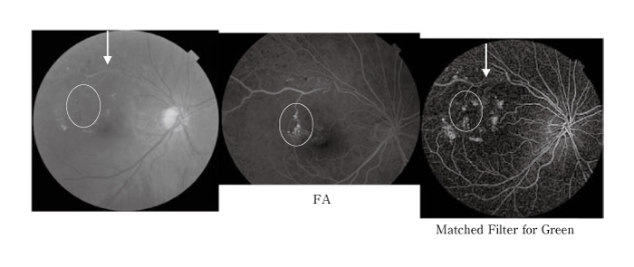
Fig. 4
Example images for old branch retinal vein occlusion; the left image was original, the middle image was for fluorescein angiogram (FA), and the right image for the matched filters. The arrows showed occluded vein, and the circles showed aneurysms. Exudates were noted for the original and the matched filters.