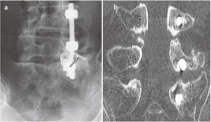
Fig. 5
Selective radiculography (a) and CT after selective radiculography (b) after surgery demonstrated that the L5 spinal nerve was not compressed between the transverse process and the sacral alar (arrows).
Fig. 5
Selective radiculography (a) and CT after selective radiculography (b) after surgery demonstrated that the L5 spinal nerve was not compressed between the transverse process and the sacral alar (arrows).